

Assessment and Quantification of Foam Cells and Lipid Droplet–Accumulating Microglia in Mouse Brain Tissue Using BODIPY Staining
使用BODIPY染色评估和定量小鼠脑组织中的泡沫细胞和脂滴累积小胶质细胞
发布: 2024年11月05日第14卷第21期 DOI: 10.21769/BioProtoc.5107 浏览次数: 2381
评审: Anonymous reviewer(s)
参见作者原研究论文
The authors used this protocol in:
Nov 2023
Advertisement


Abstract
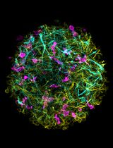
This paper presents a refined, user-friendly protocol for using boron-dipyrromethene (BODIPY) to assess and quantify foam cells and lipid droplet–accumulating microglia (LDAM) in mouse brain tissue. The protocol aims to enhance existing methodologies by offering precise and efficient evaluation of foam cells and LDAM burden in various neuropathological conditions linked to lipid metabolism and neuroinflammation. A notable challenge in analyzing tissue from mouse models of these neurodegenerative disorders is the interference caused by the autofluorescent molecule lipofuscin. Our protocol addresses this issue with specific steps that effectively distinguish BODIPY fluorescence from lipofuscin autofluorescence, using advanced imaging techniques and filter settings to ensure accurate and reliable analysis. By providing a straightforward and accessible method, this research aims to facilitate the broader adoption of BODIPY-based techniques for detailed foam cell and LDAM analysis in mouse brain tissue, potentially enhancing diagnostic capabilities and deepening our understanding of how these cells contribute to neurodegenerative disease mechanisms.
Key features
• To induce foam cell/LDAM CNS formation, this protocol was developed using brain tissue from mice subjected to permanent occlusion of the middle cerebral artery.
• The protocol utilizes mouse brain tissue that is fixed in 4% PFA.
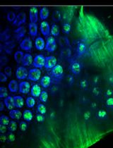
• Additional markers, CD68 and Iba1, are incorporated to evaluate myeloid cell lineage.
• The protocol includes a simple method for distinguishing BODIPY fluorescence from autofluorescence.
Keywords: Foam cells (泡沫细胞)Background
Foam cell accumulation, resulting from the overwhelmed processing of myelin-derived lipids by macrophages and microglia, plays a pivotal role in multiple pathological conditions affecting the central nervous system (CNS), including stroke, spinal cord injury, and multiple sclerosis (MS) [1–3]. In the aging brain, lipid droplet–accumulating microglia (LDAM) signify a dysfunctional and pro-inflammatory state [4]. However, myeloid cells that accumulate lipid droplets often exhibit autofluorescence due to the presence of lipofuscin, complicating accurate visualization and quantification. Currently, there is a lack of user-friendly assessment protocols for accurately measuring the burden of foam cells and LDAM in mouse models of neurological disorders. The goal of this paper is to bridge this gap by introducing a comprehensive and accessible boron-dipyrromethene (BODIPY)-based protocol tailored for the analysis of foam cells and LDAM in highly autofluorescent mouse brain tissue.
The BODIPY-based approach outlined in this study leverages the distinctive properties of BODIPY dyes, especially BODIPY 493/503, for specific neutral lipid staining and efficient visualization of foam cells. Particularly well-suited for fluorescence microscopy, this technique offers a level of detail unmatched by alternative methods like Oil Red O or H&E staining. The BODIPY-based staining method we describe is rapid, straightforward, and cost-effective. Additionally, BODIPY staining can be combined with immunostaining for a more comprehensive analysis of myeloid cell phenotype. However, despite the relatively narrow emission spectrum of BODIPY 493/503, peaking at around 503 nm, careful deployment is essential to mitigate interference from the autofluorescent molecule lipofuscin, which has a heterogeneous emission spectrum ranging from 480 to 695 nm [5]. This creates the potential for spectral overlap, even though the emission spectra do not perfectly align.
By presenting a protocol that overcomes the limitations posed by lipofuscin autofluorescence, this study aims to empower researchers with a robust and user-friendly fluorescent tool for studying foam cell and lipid droplet load in mouse brain tissue. The accessibility of this BODIPY-based protocol may encourage broader adoption and implementation, thereby promoting advancements in our understanding of the relationship between foam cells and LDAM accumulation in the aging CNS and various neurological disorders.
Materials and reagents
Biological materials
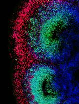
Tissue sections: 40 µm thick sections from saline perfused C57BL/6 mice (Jackson Laboratories, catalog number: 00064), fixed in 4% paraformaldehyde for 24 h and cryopreserved in 30% sucrose using standard techniques. These mice were sacrificed seven weeks post a distal middle cerebral artery occlusion model of stroke, as described by Doyle et al. [6]
Reagents
Rat anti-mouse CD68 (Bio-Rad, catalog number: MCA1957GA)
Rabbit anti-mouse Iba1 (FUJIFILM Wako Pure Chemical Corporation, catalog number: 019-19741)
Goat anti-rabbit IgG (H+L) cross-adsorbed secondary antibody, Alexa FluorTM 568 (Thermo Fisher Scientific, catalog number: A-11011)
Goat anti-rat IgG (H+L) cross-adsorbed secondary antibody, Alexa FluorTM 647 (Thermo Fisher Scientific, catalog number: A-21247)
BODIPY 493/503 (4,4-Difluoro-1,3,5,7,8-Pentamethyl-4-Bora-3a,4a-Diaza-s-Indacene) (Thermo Fisher Scientific, catalog number: D3922)
Dimethyl sulfoxide (DMSO) (Thermo Fisher Scientific, Gibco®, catalog number:25200-056)
Antifade mounting medium (Vector Laboratories, SKU: H-1400-10)
10× PBS buffer pH 7.4 (Thermo Fisher Scientific, catalog number: AM9625)
Goat serum (Atlanta Biologicals, catalog number: S131x)
Triton X-100 (MilliporeSigma, catalog number: 9002-93-1)
Solutions
0.1 M PBS (see Recipes)
0.3% Triton X-100 in 0.1 M PBS (see Recipes)
Recipes
0.1 M PBS
Reagent Final concentration Amount 10× PBS 10% 100 mL Double-distilled H2O 90% 900 mL Total 10% 1,000 mL 0.3% Triton X-100 in 0.1 M PBS
Reagent Final concentration Amount 0.1 M PBS 99.7% 498.5 mL Triton X-100 0.3% 1.5 mL Total 0.3% 1,000 mL
Laboratory supplies
12-well tissue culture plate (CELLTREAT Scientific Products, catalog number: 229112)
Brushes for tissue handling (Precisionary, SKU: VF-VM-PB-CANAL)
Hydrophobic barrier pap pen (Thermo Fisher Scientific, catalog number: R3777)
Round cover glass, #1.5 thickness, 12 mm, 100 pack (Fisher Scientific, Thomas Scientific, catalog number: NC1129240)
Superfrost PlusTM microscope slides white tab (Fisher Scientific, Fisherbrand, catalog number: 1255015)
Micro-centrifuge transfer pipette (RPI Research Products International, SKU: 147500)
15 mL conical centrifuge tubes (Fisher Scientific, FalconTM, catalog number: 14-959-53A)
10 μL micropipette tips (USA Scientific, catalog number: 1161-3700)
200 μL micropipette tips (USA Scientific, catalog number: 1163-1700)
1,250 μL micropipette tips (USA Scientific, catalog number: 1161-1820)
Equipment
Digital orbital shaker (LabniqueTM, catalog number: MT-201-BX)
Leica DM6000B microscope (Leica Microsystems, SKU: 14628696)
Zeiss LSM880 NLO upright multiphoton/confocal microscope
Software and datasets
ImageJ (free, https://imagej.net/software/fiji/downloads, Fiji version, 2024)
Procedure
文章信息
稿件历史记录
提交日期: Jun 28, 2024
接收日期: Sep 9, 2024
在线发布日期: Oct 15, 2024
出版日期: Nov 5, 2024
版权信息
© 2024 The Author(s); This is an open access article under the CC BY license (https://creativecommons.org/licenses/by/4.0/).
如何引用
Maiyo, B. K., Loppi, S. H., Morrison, H. W. and Doyle, K. P. (2024). Assessment and Quantification of Foam Cells and Lipid Droplet–Accumulating Microglia in Mouse Brain Tissue Using BODIPY Staining. Bio-protocol 14(21): e5107. DOI: 10.21769/BioProtoc.5107.
分类
神经科学 > 细胞机理 > 小神经胶质
细胞生物学 > 细胞成像 > 荧光
您对这篇实验方法有问题吗?
在此处发布您的问题,我们将邀请本文作者来回答。同时,我们会将您的问题发布到Bio-protocol Exchange,以便寻求社区成员的帮助。