

Quantification of Macrophage Cellular Ferrous Iron (Fe2+) Content using a Highly Specific Fluorescent Probe in a Plate-Reader
在平板检测仪中使用高特异性荧光探针定量巨噬细胞细胞二价铁 (Fe2+) 含量
发布: 2024年02月05日第14卷第3期 DOI: 10.21769/BioProtoc.4929 浏览次数: 2226
评审: Alka MehraAnonymous reviewer(s)
参见作者原研究论文
The authors used this protocol in:

Aug 2023
Advertisement


Abstract
Macrophages are at the center of innate immunity and iron metabolism. In the case of an infection, macrophages adapt their cellular iron metabolism to deprive iron from invading bacteria to combat intracellular bacterial proliferation. A concise evaluation of the cellular iron content upon an infection with bacterial pathogens and diverse cellular stimuli is necessary to identify underlying mechanisms concerning iron homeostasis in macrophages. For the characterization of cellular iron levels during infection, we established an in vitro infection model where the murine macrophage cell line J774A.1 is infected with Salmonella enterica serovar Typhimurium (S.tm), the mouse counterpart to S. enterica serovar Typhi, under normal and iron-overload conditions using ferric chloride (FeCl3) treatment. To evaluate the effect of infection and iron stimulation on cellular iron levels, the macrophages are stained with FerroOrange. This fluorescent probe specifically detects Fe2+ ions and its fluorescence can be quantified photometrically in a plate reader. Importantly, FerroOrange fluorescence does not increase with chelated iron or other bivalent metal ions. In this protocol, we present a simple and reliable method to quantify cellular Fe2+ levels in cultured macrophages by applying a highly specific fluorescence probe (FerroOrange) in a TECAN Spark microplate reader. Compared to already established techniques, our protocol allows assessing cellular iron levels in innate immune cells without the use of radioactive iron isotopes or extensive sample preparation, exposing the cells to stress.
Key features
• Easy quantification of Fe2+ in cultured macrophages with a fluorescent probe.
• Analysis of iron in living cells without the need for fixation.
• Performed on a plate reader capable of 540 nm excitation and 585 nm emission by trained employees for handling biosafety level 2 bacteria.
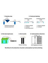
Graphical overview

Background
Human typhoid fever, a severe and often life-threatening infectious disease, is caused by the facultative intracellular Gram-negative bacterium Salmonella enterica serovar Typhi, leading to major health loss globally (Stanaway et al., 2019). For murine infection models, the strain Salmonella enterica serovar Typhimurium (Salmonella Typhimurium, S.tm), which causes a systemic disease in mice but a self-limiting gastroenteritis in humans, is frequently used.
In the case of a bacterial infection, invading pathogens are phagocytized by macrophages, the first line of innate immune defense (Weiss and Schaible, 2015). S.tm, however, thrives within macrophages. Despite its ability to invade various types of cells, virulence is dependent on intramacrophage proliferation (Fields et al., 1986; Leung and Finlay, 1991). One factor critically affecting the outcome of this host–pathogen interaction is the availability of nutrients, as intramacrophage S.tm depends on the acquisition of essential nutrients to sustain efficient proliferation. This includes amino acids or trace metals, like iron. On one hand, S.tm–driven metabolic reprogramming grants the pathogen access to intracellular nutrients (Liss et al., 2017). On the other hand, macrophages exploit this nutrient demand by withdrawing iron from the spatio-temporal localization of the pathogen in a process termed nutritional immunity (Nairz et al., 2007; Murdoch and Skaar, 2022). Appropriately, a state of systemic or cellular iron excess is associated with increased bacterial proliferation (Khan et al., 2007; Porto and De Sousa, 2007; Kao et al., 2016).
Macrophage cellular iron metabolism and its adaptation to an infection have been extensively studied. In the case of an infection with an intracellular pathogen like S.tm, regulation of the key iron metabolism proteins Ferroportin-1 (FPN1) and the Transferrin receptor-1 (TFR1) facilitates a decrease of cellular iron content and thus leads to an improved infection control of intracellular bacteria (Nairz et al., 2008; Fritsche et al., 2012; Wessling-Resnick, 2015; Abreu et al., 2020). Furthermore, transport of iron into the cytosolic lumen is accomplished by the divalent metal transporter-1 (DMT1) and by the natural resistance-associated macrophage protein-1 (NRAMP1 or SLC11A1), both of which have also been implicated in bacterial iron withdrawal (Forbes and Gros, 2003; Fritsche et al., 2012; Grander et al., 2022). DMT1 is responsible for the uptake of non-transferrin-bound iron (NTBI) from outside the macrophage and for transporting transferrin-bound iron (TBI) from the early endosome into the lumen of the phagocyte. NRAMP1 transports iron out of the late phagosome. As both are only capable of binding Fe2+, the function of the six-transmembrane epithelial antigen of prostate 3 (STEAP3) in the late endosome to reduce Fe3+ to Fe2+ is indispensable (Wang and Pantopoulos, 2011).
Another factor at play is the acute phase protein hepcidin (HAMP1), a liver-derived hormone regarded as the systematic master regulator of iron metabolism. During an infection, hepcidin targets the iron exporter FPN1, leading to its degradation and thus iron sequestration, causing hypoferremia (Nemeth et al., 2004). However, its role in an infection with intracellular pathogens is not yet completely understood (Chlosta et al., 2006; Lim et al., 2018).
During infection studies, cellular iron quantification is frequently relevant. Standard procedures like the usage of radioactive 59Fe isotopes are elaborate, expensive, and might be inapplicable in certain experimental settings. Iron quantification with the help of the quenchable probe Calcein-AM is easily available and often used but has several disadvantages. Acquiring fluorescence by flow cytometry needs extensive preparation of samples that exposes cells to mechanical stress; furthermore, as Calcein does not pass into the cellular membrane compartments (e.g., lysosomes), which are rich in labile iron (chelatable), total cellular iron is most likely drastically underestimated when this method is applied (Tenopoulou et al., 2007).
Herein, we report a simple and powerful tool to accurately determine alterations in cellular iron levels upon diverse stimuli, which can be employed for cultured immune cells, here exemplified in a murine macrophage infection with intracellular bacteria. As the cellular iron trafficking machinery primarily utilizes Fe2+, monitoring metabolically active intracellular ferrous iron levels is most insightful (Hentze et al., 2010; Moroishi et al., 2011; Cronin et al., 2019). We apply a fluorescent probe that specifically detects Fe2+, based on N-oxide chemistry (RhoNox-4; commercial name: FerroOrange) (Hirayama et al., 2020). By using this approach, cellular levels of the trace metal can be quantified in a plate reader without additionally exposing cells to stressors.
Materials and reagents
12-well plate (Falcon, catalog number: 353043)
Acridine orange/propidium iodide stain (Biocat, catalog number: F23001)
Agar-Agar Kobe I (Roth, catalog number: 5210.3)
Aqua bidest (Fresenius Kabi, catalog number: 16.231)
CASY Cup (OMNI Life Science, catalog number: 5651794)
CASY Ton buffer (OMNI Life Science, catalog number: 5651808)
Cell scraper (Sarstedt, catalog number: 83.3951)
CoolCellTM LX freezing container (Merck, catalog number: BCS-405G)
Cryo vial with silicone washer, 2 mL (Simport, catalog number: T311-3)
Dimethylsulfoxide (DMSO) (Roth, catalog number: A994.1)
Disposable cuvette (BRAND, catalog number: 759015)
Disposable pipettes, 5 mL, 10 mL, and 25 mL (Falcon, catalog number: 606180, 607180, and 357525, respectively)
Dulbecco’s modified Eagle’s medium (DMEM) (Pan BiotechTM, catalog number: P04-01500)
Eppendorf tubes, 0.5 mL (Eppendorf, catalog number: 0030121.023)
Erlenmeyer flask, 250 mL (Stoelzle Medical, catalog number: 21226368000)
Ferric chloride (FeCl3) (Sigma, catalog number: 236489)
FerroOrange (GERBU Biotechnik GmbH, catalog number: F374-10)
Fetal bovine serum (FBS) (Pan BiotechTM, catalog number: P30-3031)
Gentamicin (Gibco, catalog number: 15750-037), stock: 50 mg/mL
Glass beaker, 150 mL (Ruprechter, catalog number: 102113729)
Glycerol (Sigma, catalog number: G5516-100ML)
Iscove’s modified Dulbecco's medium (IMDM) (Pan BiotechTM, catalog number: P04-20150S3)
L-Glutamine (Lonza, catalog number: BE17-605E)
Luna cell counting slides (Biocat, catalog number: L201B1C3GB)
Lysogeny broth (LB) medium Lennox (Roth, catalog number: X964.2)
Penicillin/streptomycin (Capricorn Scientific, catalog number: PS-B)
Phosphate buffer saline (PBS) (Lonza, catalog number: 17-515 F)
Pipetman L Starter Kit, 2, 20, 200, and 1,000 μL pipettes (GILSON, catalog number: F167370)
Pipette tips, 10, 200, and 1,250 μL (STARLAB, catalog number: S1110-3700, S1120-3810, and S1112-1720, respectively)
Polypropylene tube, 50 mL (Falcon, catalog number: 352070)
Salmonella enterica serovar Typhimurium ATCC14028 (ATCC)
Sartorius Midi Plus pipetting controller (Sartorius, catalog number: 710931)
Tissue culture flask, 750 mL, straight neck (Falcon, catalog number: 353028)
LB medium (see Recipes)
LB medium with 30% glycerol (see Recipes)
Cell culture medium (see Recipes)
Cell culture medium for infection with S.tm (see Recipes)
Staining solution (see Recipes)
Iron (III) chloride solution (see Recipes)
Cell line
J774A.1 (ATCC TIB-67TM) is a macrophage cell line isolated in 1968 from a female BALB/c mouse with reticulum cell sarcoma.
Recipes
LB medium
200 mL aqua bidest
2 g LB medium powder
Autoclave (20 min at 121 °C and 10 min at 50 °C)
LB medium with 30% glycerol
Add 300 μL of glycerol to 700 μL of LB medium
Cell culture medium
500 mL of DMEM
50 mL of FBS
5 mL of L-Glutamine
5 mL of Penicillin/Streptomycin
Cell culture medium for infection with S.tm
500 mL of DMEM
5 mL of FBS
5 mL of L-Glutamine
Staining solution
5 mL of IMDM
1 μL of FerroOrange (1 mmol/L)
Iron (III) chloride solution
135 mg of FeCl3
50 mL of Aqua bidest
Equipment
Automated multimode microplate reader (TECAN Spark, catalog number: 1912001805)
Casy counting system CASY TT (OMNI Life Science, catalog number: TT2QA2571)
Centrifuge (Hettich Micro 200R and Rotanta 460R)
Freezer, -80 °C (Thermo Fisher Scientific, catalog number: 15788587)
CO2 incubator (Thermo Fisher Scientific, model: Heraeus® HERAcell®)
Laminar flow cabinet (EuroClone Sicherheitswerkbank Safe Mate Eco 1.2) (Politakis Laborgeräte, catalog number: EN 12 469)
Liquid nitrogen storage tank (CryoShop, catalog number: CS-79105601)
LUNA automated cell counter (Biocat, catalog number: L10001-LG)
Millivac-Maxi vacuum pump (Merck, catalog number: SD1P014M04)
Photometer (Eppendorf, catalog number: BioPhotometer D30)
Shaking incubator (VWR, catalog number: GFL 3031)
Software and datasets
GraphPad Prism 9.1 (GraphPad Software)
SparkControlTM (Spark Method Editor V.3, release date 2021 06 01) (TECAN Trading, Ltd.)
Procedure
文章信息
版权信息
© 2024 The Author(s); This is an open access article under the CC BY-NC license (https://creativecommons.org/licenses/by-nc/4.0/).
如何引用
Grubwieser, P., Brigo, N., Seifert, M., Grander, M., Theurl, I., Nairz, M., Weiss, G. and Pfeifhofer-Obermair, C. (2024). Quantification of Macrophage Cellular Ferrous Iron (Fe2+) Content using a Highly Specific Fluorescent Probe in a Plate-Reader. Bio-protocol 14(3): e4929. DOI: 10.21769/BioProtoc.4929.
分类
免疫学 > 免疫细胞功能 > 巨噬细胞
细胞生物学 > 细胞新陈代谢 > 其它化合物
您对这篇实验方法有问题吗?
在此处发布您的问题,我们将邀请本文作者来回答。同时,我们会将您的问题发布到Bio-protocol Exchange,以便寻求社区成员的帮助。