

Studying Cell Migration (Random and Wound Healing) Parameters with Imaging and MATLAB Analysis
利用成像和MATLAB分析研究细胞迁移(随机和伤口愈合)参数
(*contributed equally to this work) 发布: 2023年11月05日第13卷第21期 DOI: 10.21769/BioProtoc.4871 浏览次数: 1544
评审: Alka MehraAnaïs PanosaAnonymous reviewer(s)
参见作者原研究论文
The authors used this protocol in:
Jul 2021
Advertisement


Abstract
Cell migration is an essential biological process for organisms, in processes including embryonic development, immune response, and cancer metastasis. To elucidate the regulatory machinery of this vital process, methods that mimic in vivo migration, including in vitro wound healing assay and random migration assay, are widely used for cell behavior investigation. However, several concerns are raised with traditional cell migration experiment analysis. First, a manually scratched wound often presents irregular edges, causing the speed analysis difficult. Second, only the migration speed of leading cells is considered in the wound healing assay. Here, we provide a reliable analysis method to trace each cell in the time-lapse images, eliminating the concern about wound shape and creating a more comprehensive understanding of cell migration—not only of collective migration speed but also single-cell directionality and coordination between cells.
Keywords: Live-cell imaging (活细胞成像)Background
Cell migration is an individual or collective cell movement that plays an important role in embryonic development (Weijer, 2009), immune response (Luster et al., 2005), and cancer metastasis (Friedl and Glimour, 2009). To investigate these essential physiological phenomena in vitro, wound healing assay or random migration assay are often applied (Jin et al., 2016).
Compared with traditional analyses of wound healing assay focusing on general migration speed, our script mainly focuses on precisely tracking each cell in each timeframe image, leading to speed, directionality, and coordination being analyzed simultaneously. Moreover, we can conquer the difficulties in analyzing the wound healing assay even when the edge of the wound is not smooth.
Since wound healing assay provides cell information like migration speed and directionality, and random migration provides cell coordination information, we applied both assays to elucidate the mechanisms of cell migration thoroughly. We found several pairs of genes and pathways that affect migration speed and cell coordination through these assays after performing two-hit inhibition: the knockdown of a migration-related gene using shRNA combined with the addition of an inhibitor of migration-related pathways at the same time. One of our best candidates, STK40 and MAPK, showed a synergistic effect on migration speed. By applying shSTK40 and MAPK inhibitor PD98059, we found that the absence of either of them has a mild effect on cell migration speed, while the speed significantly dropped when both of them were inhibited at the same time.
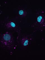
Our method is a half-automatic way to trace single cells in the time-lapse immunofluorescence images, which provides multiple parameters for a comprehensive analysis of cell migration, including speed, density, directionality, and cell–cell coordination. In sum, our method not only solves the aforementioned concern but provides a fast, cheap, and comprehensive analysis of cell migration.
Materials and reagents
96-well black/clear-bottom plate (Thermo Fisher Scientific, NuncTM, catalog number: 165305)
Collagen I, rat tail (Thermo Fisher Scientific, GibcoTM, catalog number: A1048301)
Hoechst 33342 (Thermo Fisher Scientific, InvitrogenTM, catalog number: H3570)
HEPES (Thermo Fisher Scientific, GibcoTM, catalog number: 15630106)
Bovine serum albumin (BSA) (BioShop, catalog number: ALB001)
Recombinant human EGF (PeproTech, catalog number: AF-100-15)
Human FGF-acidic recombinant protein (Thermo Fisher Scientific, GibcoTM, catalog number: PHG0014)
Heparin sodium salt from porcine intestinal mucosa (Merck, Sigma-Aldrich, catalog number: H3393)
Trametinib (LC laboratories, catalog number: T-8123)
PBS, pH 7.4 (Thermo Fisher Scientific, GibcoTM, catalog number: 10010023)
Cell migration supplements (see Recipes)
Cell lines
SAS cell line was used as indicated. Cells were grown in Dulbecco’s modified Eagle medium (DMEM) with 10% fetal bovine serum (FBS), and 1% penicillin and streptomycin.
Recipes
Cell migration supplements
20 mM HEPES
0.1% BSA
5 ng/mL EGF or 25 ng/mL FGF1
10 U heparin
Equipment
Fluorescence microscope (Nikon Eclipse Ti)
96-well scratcher (can be replaced by 200 μL tip or toothpick)
Software
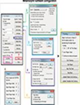
MATLAB (MathWorks, https://www.mathworks.com/products/matlab.html) with the image processing toolbox (access date, 8/1/2017)
Nikon NIS-Elements Viewer (Nikon image acquisition software) (access date, 9/21/2017)
Fiji from ImageJ (open resource software for image analysis) (access date, 8/1/2017)
Excel (Microsoft) (access date, 8/1/2017)
Procedure
文章信息
版权信息
© 2023 The Author(s); This is an open access article under the CC BY-NC license (https://creativecommons.org/licenses/by-nc/4.0/).
如何引用
Readers should cite both the Bio-protocol article and the original research article where this protocol was used:
- Yu, L. Y., Lin, H. C., Hsu, C. L., Kao, T. Y. and Tsai, F. C. (2023). Studying Cell Migration (Random and Wound Healing) Parameters with Imaging and MATLAB Analysis. Bio-protocol 13(21): e4871. DOI: 10.21769/BioProtoc.4871.
- Yu, L. Y., Tseng, T. J., Lin, H. C., Hsu, C. L., Lu, T. X., Tsai, C. J., Lin, Y. C., Chu, I., Peng, C. T., Chen, H. J., et al. (2021). Synthetic dysmobility screen unveils an integrated STK40-YAP-MAPK system driving cell migration. Sci. Adv. 7(31): eabg2106.
分类
癌症生物学 > 侵袭和转移 > 细胞生物学试验 > 细胞迁移
细胞生物学 > 细胞信号传导 > 胞内信号传导
您对这篇实验方法有问题吗?
在此处发布您的问题,我们将邀请本文作者来回答。同时,我们会将您的问题发布到Bio-protocol Exchange,以便寻求社区成员的帮助。