

Mouse Corneal Epithelial and Stromal Cell Isolation and Culture
小鼠角膜上皮和基质细胞的分离和培养
发布: 2023年10月05日第13卷第19期 DOI: 10.21769/BioProtoc.4829 浏览次数: 2928
评审: Pilar Villacampa AlcubierreMarina Sánchez PetidierAnonymous reviewer(s)
参见作者原研究论文
The authors used this protocol in:

Apr 2023
Advertisement


Abstract
Corneal epithelium and stroma are the major cellular structures for ocular protection and vision accuracy; they play important roles in corneal wound healing and inflammation under pathological conditions. Unlike human, murine corneal and stromal fibroblast cells are difficult to isolate for cell culture. In our laboratory, we successfully used an ex vivo culture procedure and an enzymatic procedure to isolate, purify, and culture mouse corneal epithelial and stromal fibroblast cells.
Key features
• Primary cell culture models of a disease are critical for cellular and molecular mechanism studies.
• Corneal tissues with the limbus contain stem cells to generate both epithelial and stromal cells.
• An ex vivo corneal culture provides a constant generation of primary corneal cells for multiple passages.
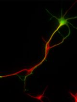
• The isolated cells are validated by the corneal epithelial cell markers Krt12 and Cdh1 and the stromal fibroblast marker Vim.
Keywords: Mouse cornea (小鼠角膜)Background
The cornea is a transparent avascular tissue; it acts as a barrier to protect the eye and has a 2/3 focal power to project an image onto the retina. Mouse cornea consists of three cellular layers: the epithelium, the stroma, and the endothelium (Figure 1). The epithelium consists of a differentiated superficial squamous cell, a transit wing cell, and a proliferative basal cell type, and is the outmost ocular tissue that contains 80% corneal cells. The innermost corneal tissue is the endothelium, consisting of a single unrenewable cell sheet. The epithelium layer and the endothelium layer are separated from the stroma by the Bowman’s membrane and the Descemet’s membrane, respectively. The stroma is the thickest and toughest part of the cornea in between the epithelium and the endothelium. Although the stroma contains only approximately 20% of the corneal cells, it occupies two thirds of the corneal space. Both epithelial and stromal cells are embedded in a tough collagen-rich extracellular matrix (ECM) [1]. As the epithelial cells can be regenerated by a group of stem cells, also known as limbal epithelial stem cells, at the limbus (the border area between the cornea and the sclera) for cellular homeostasis and wound repair, it is relatively easy to isolate and culture them [2]. However, the corneal stromal fibroblast (keratocyte) may be regenerated at a relatively slower pace; its regeneration is supposed to be either local and/or come from the progenitor at the limbus region. Isolation and culture of corneal cells, i.e., epithelial cells and stromal keratocytes, are critical for the study of corneal diseases, particularly wound healing [3] and neovascularization [4], as well as their underlying molecular mechanisms [5]. Human corneal primary epithelial and stromal cells and murine primary corneal epithelial cells are commercially available; however, no murine primary stromal fibroblast cells can be purchased. It is difficult to isolate and culture murine primary corneal cells, particularly murine keratocytes. A protocol to isolate and culture murine keratocytes is available for newborn pups, where the separation of the stroma from the epithelium is very difficult and the resultant cell identification was not verified by a specific marker [6]. We have developed a relatively simple and reliable method to isolate both murine corneal epithelial cells and keratocytes, and culture them in vitro for multiple passages.

Figure 1. Mouse corneal structure and cell types
Materials and reagents
Biological materials
Animals: 4–8-week-old B6 mice (The Jackson Laboratory, catalog number: 000664)
Consumable materials
Cell culture plate: 12-well cell culture plates (Thermo Fisher Scientific, catalog number: 150628)
Eppendorf tubes (Thermo Fisher Scientific, catalog number: 3451)
Tipone® pipette tips (USA Scientific, catalog number: 1126-7810)
Pipettes (Corning, Falcon®, catalog number: 357543)
Cryopreservation vials (Corning, catalog number: 430659)
Reagents
Ethanol
Phosphate-buffered saline (PBS) (Sigma-Aldrich, catalog number: P4417-100TAB)
Penicillin-streptomycin (5,000 U/mL) (Thermo Fisher Scientific, catalog number: 15070063)
Dulbecco’s modified Eagle’s medium (DMEM, high glucose) (Thermo Fisher Scientific, catalog number: 11965)
Fetal bovine serum (FBS) (GE Healthcare, HyClone, catalog number: SH30071.03)
Gelatin (Sigma-Aldrich, catalog number: G1890-100G)
Collagenase A (Millipore Sigma, catalog number: 10103578001)
Trypsin ethylenediaminetetraacetic acid (EDTA) solution (Mediatech, Cellgro®, catalog number: 25-053-CI)
Common epithelial marker E-cadherin (Cdh1) (BD Transduction Lab, catalog number: 610181)
Specific epithelial marker Keratin 12 (Krt12) (Abclonal, catalog number: A9642)
Fibroblast marker vimentin (Sigma, catalog number: 17-10195)
100× penicillin-streptomycin solution (Thermo Fisher Scientific, catalog number: 15140122)
Corneal epithelial cell basal medium (ATCC, catalog number: PCS-700-030)
Corneal epithelial cell growth kit (ATCC, catalog number: PCS-700-040)
Solutions
Corneal epithelial cell culture medium (see Recipes)
Corneal stromal fibroblast culture medium (see Recipes)
0.1% gelatin solution (see Recipes)
Recipes
Corneal epithelial cell culture medium
Prepare the complete corneal epithelial growth medium according to the manufacturer’s instructions. The final concentration for each component in ATCC complete corneal epithelial growth medium is as follows:
5 μg/mL Apo-transferrin
1.0 μM epinephrine
0.4% extract P
100 ng/mL hydrocortisone
6 mM L-glutamine
5 μg/mL recombinant human insulin
CE Growth Factor: proprietary formulation
Corneal stromal fibroblast culture medium
450 mL of DMEM
50 mL of FBS
5 mL of penicillin/streptomycin (5,000 U/mL)
0.1% gelatin solution
0.1 g gelatin powder
100 mL of PBS
Autoclave and store at 4 °C for three months.
Equipment
Mcpherson-Vannas curved iris scissors (Storz Ophthalmic Instruments, catalog number: E3347)
Castroviejo suturing forceps 0.12 mm (Storz Ophthalmic Instruments, catalog number: E1796)
Martinez double ended corneal dissector (Storz Ophthalmic Instruments, catalog number: E3001)
Algerbrush II (Katena Products, Inc., Denville, NJ)
Cell culture incubator (Thermo Fisher Scientific, Thermo Scientific, model: 3250)
Water bath (VWR, model: 1545)
Pipettes (Eppendorf)
Pipet-aid (Drummond)
Benchtop centrifuge (Beckman Coulter, model: Allegra® X-15R)
Lab rotating shaker (Barnstead Thermolyne LabQuake, model: 4152110)
Inverted microscope (Nikon, Eclipse TS100)
Confocal microscope (Nikon, Eclipse Ti)
40 μm nylon net filter (Fisher Scientific, Leicestershire, UK)
Procedure
文章信息
版权信息
© 2023 The Author(s); This is an open access article under the CC BY-NC license (https://creativecommons.org/licenses/by-nc/4.0/).
如何引用
Zhang, Y., Zhang, L. and Liu, Y. (2023). Mouse Corneal Epithelial and Stromal Cell Isolation and Culture. Bio-protocol 13(19): e4829. DOI: 10.21769/BioProtoc.4829.
分类
细胞生物学 > 细胞分离和培养 > 单层培养
生物工程 > 生物医学工程
您对这篇实验方法有问题吗?
在此处发布您的问题,我们将邀请本文作者来回答。同时,我们会将您的问题发布到Bio-protocol Exchange,以便寻求社区成员的帮助。