

Quantitative Analysis of Clot Deposition on Extracorporeal Life Support Membrane Oxygenators Using Digital and Scanning Electron Microscopy Imaging Techniques
使用数字和扫描电子显微镜成像技术对体外生命支持膜氧合器上的凝块沉积进行定量分析
发布: 2023年09月20日第13卷第18期 DOI: 10.21769/BioProtoc.4814 浏览次数: 1513
评审: Komuraiah MyakalaBhanu JagilinkiAndrew Doyle
Advertisement


Abstract
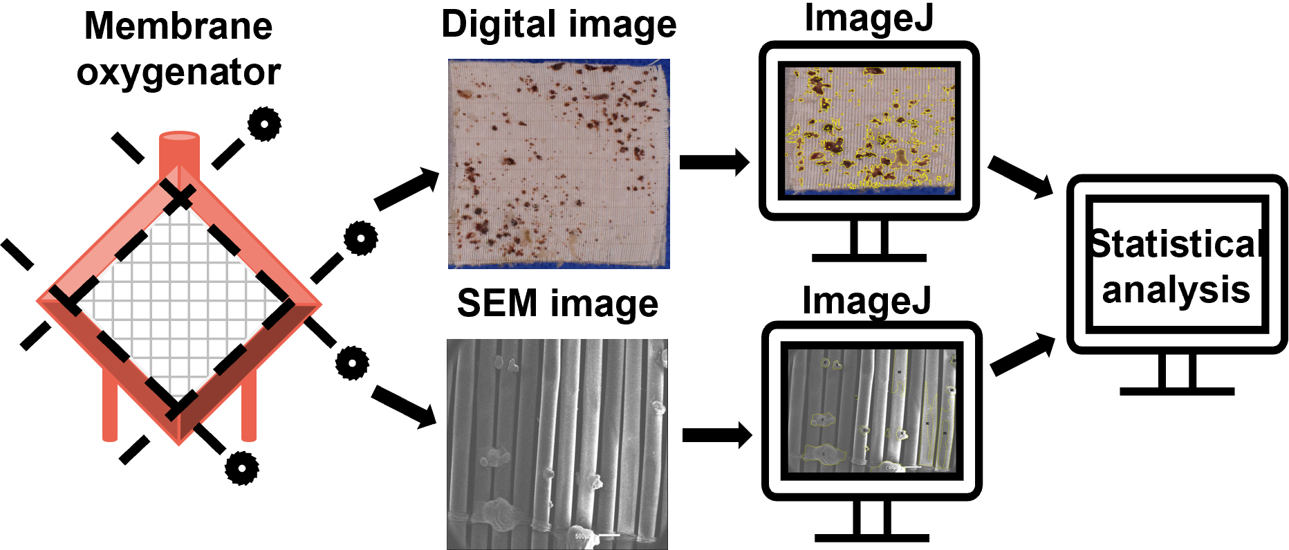
Device-induced thrombosis remains a major complication of extracorporeal life support (ECLS). To more thoroughly understand how blood components interact with the artificial surfaces of ECLS circuit components, assessment of clot deposition on these surfaces following clinical use is urgently needed. Scanning electron microscopy (SEM), which produces high-resolution images at nanoscale level, allows visualization and characterization of thrombotic deposits on ECLS circuitry. However, methodologies to increase the quantifiability of SEM analysis of ECLS circuit components have yet to be applied clinically. To address these issues, we developed a protocol to quantify clot deposition on ECLS membrane oxygenator gas transfer fiber sheets through digital and SEM imaging techniques. In this study, ECLS membrane oxygenator fiber sheets were obtained, fixed, and imaged after use. Following a standardized process, the percentage of clot deposition on both digital images and SEM images was quantified using ImageJ through blind reviews. The interrater reliability of quantitative analysis among reviewers was evaluated. Although this protocol focused on the analysis of ECLS membrane oxygenators, it is also adaptable to other components of the ECLS circuits such as catheters and tubing.
Key features
• Quantitative analysis of clot deposition using digital and scanning electron microscopy (SEM) techniques
• High-resolution images at nanoscale level
• Extracorporeal life support (ECLS) devices
• Membrane oxygenators
• Blood-contacting surfaces
Graphical overview
Background
Device-induced thrombosis remains one of the major complications of extracorporeal life support (ECLS) (Jaffer et al., 2015; Doyle and Hunt, 2018). The interaction of blood and artificial surfaces of ECLS components results in clot deposition on these surfaces and in severe instances can lead to occlusion of the circuit and systemic complications (Roberts et al., 2020a). Clinically available antithrombotic treatments can introduce hemorrhagic complications, especially in patients with preexisting bleeding disorders (MacLaren et al., 2022). Non-thrombogenic circuits that reduce thrombotic complications and enable improved patient outcomes are needed. An important prerequisite to developing such circuits is to more thoroughly understand how blood components interact with the artificial surfaces of ECLS devices (Beely et al., 2016). However, there is currently no methodology for routine examination of these devices after use. Surfaces of ECLS components could be specifically engineered to address the issue of thrombosis with a better understanding of the interactions between blood and ECLS surfaces. Examination and analysis of extracorporeal circuitry following clinical use in a standardized and validated method could inform both clinical decision-making regarding anticoagulation as well as device design.
Digital images have been used to record and visualize the extent of clot formation within ECLS devices in small pilot research studies, both clinically and in translational research laboratories (Lehle et al., 2008; Beely et al., 2016; Diehl and Gantner, 2018; Chlebowski et al., 2020; Naito et al., 2021). Blood clots are easily observed through digital images, while some microscopic thrombotic structures are not. Scanning electron microscopy (SEM), an advanced technology, produces high-resolution images at nanoscale level. Although most of the clinical use of SEM is qualitative, new methodologies that increase the quantifiability of SEM have been developed (Kundu et al., 1988; Di Iorio et al., 2005). We have previously used SEM to visualize and characterize clots and cell deposits in ECLS circuits following use in translational research studies in swine up to 72 h in duration (Beely et al., 2016; Roberts et al., 2020b). In this protocol, we developed and standardized a method to quantitatively analyze clot deposition using both digital and SEM images, allowing to routinely evaluate ECLS components following clinical use. Although this protocol focused on the analysis of ECLS membrane oxygenators only, this method could be applied to other components of the ECLS with necessary adaptations.
Materials and reagents
Reagents
Glutaraldehyde, 50% biological grade (Electron Microscopy Sciences, catalog number: 16520)
Sucrose, ≥ 99.5% (Sigma-Aldrich, catalog number: S9378)
Sodium cacodylate trihydrate, ≥ 98% (Sigma-Aldrich, catalog number: C0250)
Phosphate buffered saline (PBS) powder, 10×, pH 7.4 (Fisher BioReagents, catalog number: BP665-1)
Dehydrant alcohol, 100% (Epredia, catalog number: 6215)
Milli-Q water, 18.2 MΩ·cm, obtained through BarnsteadTM Smart2PureTM Pro Water Purification System
Solutions
Scanning electron microscopy fixative (SEM fixative) (see Recipes)
Scanning electron microscopy buffer (SEM buffer) (see Recipes)
Phosphate buffered saline, pH 7.4 (PBS) (see Recipes)
Recipes
SEM fixative
Reagent Final concentration Quantity Sucrose 0.1 M 8.5575 g Sodium cacodylate trihydrate 0.1 M 5.3508 g Glutaraldehyde, 50% 3% 15 mL Milli-Q water n/a 235 mL SEM buffer
Reagent Final concentration Quantity Sucrose 0.1 M 8.5575 g Sodium cacodylate trihydrate 0.1 M 5.3508 g Milli-Q water n/a 250 mL PBS (based on vendor instruction)
Reagent Final concentration Quantity PBS powder, 10× 1× 98.9 g Milli-Q water n/a 10 L
Laboratory supplies
Slotted tissue cassette (Epredia, catalog number: B851729WH)
Container, multi-purpose, PP, with separate snap lid, 120 mL (Globe Scientific, catalog number: 271004)
Copper conductive tape, single adhesive surface (Ted Pella, catalog number: 16072-1)
Carbon conductive tape, double coated (Ted Pella, catalog number: 16073-5)
T10 Torx Screwdriver (Tekton, model number: DST31100)
Sterilization wrap (Halyard, catalog number: 34162)
Premium blue indicating silica gel desiccant beads 3–5 mm (Dry & Dry, catalog number: X000TS1IZP)
Straight connector, female luer lock port (Qosina, catalog number: 73317)
Needleless injection site, swappable, male luer lock (Qosina, catalog number: 80147)
50 mL syringe, luer-lok tip (BD, catalog number: 309653)
Scissors
Surgical grade stainless steel forceps, nonlocking thumb handle straight delicate, serrated tips, 4 3/4" in. (McKesson, catalog number: 43-1-772)
Pathology metric ruler
Weighing paper or weighing boat
Spatula
Cylinder
Desiccator, 6"
SEM fixative/buffer waste bottle
Dehydration/ethanol waste bottle
Labels with study ID, protocol number, and date
Personal protective equipment (gloves, goggles, hearing protection, and scrubs/lab coats)
Equipment
Miter saw coupled with diamond blade (Miter saw DeWALT, model: DWS779; blade RIDGID, model: 12 in. dual-purpose)
Digital camera with lens (Nikon, camera model: D3300, lens model: AF-P DX NIKKOR 18-55 f/3.5-5.6G VR)
BarnsteadTM Smart2PureTM Pro Water Purification System (Thermo Fisher Scientific, catalog number: 50157873)
Gold sputter coater (Denton Vacuum, model: Desk II)
InTouchScopeTM scanning electron microscope (JEOL, model: JSM-IT100)
Impact suction unit (Envi health solutions, model: 326/326M)
Software and datasets
ImageJ (version: 1.53k, release date: 07/06/2021, free)
Analytics Software & Solutions (SAS) (version 9.4, release date: 07/2013, license needed)
Procedure
文章信息
版权信息
© 2023 The Author(s); This is an open access article under the CC BY-NC license (https://creativecommons.org/licenses/by-nc/4.0/).
如何引用
Zang, Y., Roberts, T. R., Harea, G. T., Beely, B. M., Olivera Perez, L. J., Ande, S., Batchinsky, M., Lee, J. H., Thrailkill, M. A., Reynolds, M. M. and Batchinsky, A. I. (2023). Quantitative Analysis of Clot Deposition on Extracorporeal Life Support Membrane Oxygenators Using Digital and Scanning Electron Microscopy Imaging Techniques. Bio-protocol 13(18): e4814. DOI: 10.21769/BioProtoc.4814.
分类
生物工程 > 生物医学工程
生物物理学 > 生物工程 > 医用生物材料
您对这篇实验方法有问题吗?
在此处发布您的问题,我们将邀请本文作者来回答。同时,我们会将您的问题发布到Bio-protocol Exchange,以便寻求社区成员的帮助。