

Preparation of Human Kidney Progenitor Cultures and Their Differentiation into Podocytes
人肾脏祖细胞培养物的制备及其向足细胞的分化
发布: 2023年08月20日第13卷第16期 DOI: 10.21769/BioProtoc.4757 浏览次数: 2282
评审: Rajesh RanjanFereshteh AzediAnonymous reviewer(s)
参见作者原研究论文
The authors used this protocol in:

Aug 2022
Advertisement


Abstract
Kidney diseases are a global health concern. Modeling of kidney disease for translational research is often challenging because of species specificities or the postmitotic status of kidney epithelial cells that make primary cultures, for example podocytes. Here, we report a protocol for preparing primary cultures of podocytes based on the isolation and in vitro propagation of immature kidney progenitor cells subsequently differentiated into mature podocytes. This protocol can be useful for studying physiology and pathophysiology of human kidney progenitors and to obtain differentiated podocytes for modeling podocytopathies and other kidney disorders involving podocytes.
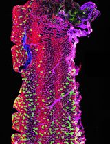
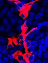
Graphical overview

Background
Kidney diseases, a global health issue, are the consequence of injury to the functional components of the kidney, the nephrons (Romagnani et al., 2017). Nephrons are constituted by a blood filtering unit, the glomerulus, and the respective tubule where the filtrate is modified by solute reabsorption and metabolite secretion up to when the final urine is excreted via the urinary tract (Romagnani et al., 2017). The nephrons respond to injury in two ways: a) differentiated epithelial cells undergo polyploidization and hypertrophy to rapidly support residual kidney function and b) immature epithelial cells, referred to as kidney progenitors (Lazzeri et al., 2019), proliferate to recover at least a part of the lost cells, i.e., kidney regeneration (Lazzeri et al., 2019). Kidney progenitors are localized along the inside of the Bowman capsule of the glomerulus and are scattered among tubular epithelial cells along the tubule, being identified by the expression of the surface markers CD133 and CD24, in humans (Sagrinati et al., 2006; Lazzeri et al., 2019). Kidney progenitors can be obtained from kidney tissue or urine and cultured long term (Angelotti et al., 2012) because they retain the capacity for self-renewal. Kidney progenitors have the capacity to differentiate into multiple types of kidney epithelial cells in vitro and in vivo (Sagrinati et al., 2006; Lazzeri et al., 2019). Hence, kidney progenitors can be expanded and differentiated into different types of tubular epithelial cells (Angelotti et al., 2012) and even cultured in 3D, generating tubuloids, a selective property among other kidney tubular cells (Xu et al., 2022). This property makes them ideal for modeling of genetic tubular disorders, e.g., upon isolation from the urine of patients with genetic tubular disorders or upon introduction of pathogenic genetic variants (e.g., using CRISPR-Cas system) (Xu et al., 2022). In addition, kidney progenitors can be differentiated in culture into podocytes, the main constituent of the glomerular filtration barrier. Podocytes are highly differentiated postmitotic cells unable to proliferate (Kopp et al., 2020). For this reason, they are impossible to expand in primary cultures, unless using artificial systems of immortalization (Shankland et al., 2007).
Here, we report detailed protocols on how to prepare human kidney progenitor cultures from human kidney tissue, maintain them, and differentiate them into podocytes. Differentiation of kidney progenitors using specific factors and compounds (Lasagni et al., 2015), as recently reported for the histone deacetylase inhibitor panobinostat, induces a change in their phenotype, promoting transcription of podocyte genes such as nephrin, podocin, and synaptopodin (Melica et al., 2022). We also report methods to assess their phenotype by qRT-PCR, FACS, and confocal and stimulated emission depletion (STED) microscopy. Applying the same culturing method described here to the isolation procedure reported by Lazzeri et al. (2015) permits the preparation of kidney progenitor cultures also from the urine of patients with kidney disorders, making them particularly suitable for studying genetic podocytopathies for diagnostic purposes. Given the importance of kidney progenitors and podocytes in the pathogenesis of chronic kidney disease, the possibility to prepare and maintain these cultures has wide implications and possible uses.
Materials and reagents
Biological materials
Normal-appearing kidney fragments are obtained from the pole opposite to the tumor from patients that underwent nephrectomy for localized renal tumors.
CAUTION: All the experiments involving human specimens should be performed in accordance with the recommendations of the Institutional Ethical Committee for human experimentation. All procedures in this protocol were conducted under protocols approved by the Ethical Committee on human experimentation of the Careggi University Hospital.
Reagents
Physiological saline solution, NaCl 0.9% (B. Braun Melsungen AG, A.I.C. n. 030902391)
HyClone defined fetal bovine serum (FBS), US origin (Cytiva, catalog number: SH30070.03)
Endothelial cell growth basal medium (EBM), 500 mL (LONZA, catalog number: CC-3121)
Microvascular endothelial growth medium (EGM-MV) SingleQuots kit (LONZA, catalog number: CC-4123) [contains growth factors, cytokines, and supplements: bovine brain extract w/o heparin (BBE); hydrocortisone (hEGF); gentamicin, amphotericin B (GA-1000); and FBS]
DMSO (Merck KGaA, catalog number: D8418)
Dulbecco’s modified Eagle’s medium/Ham’s nutrient mixture F-12 (DMEM-F12-1L) (Merck KGaA, catalog number: D2906)
Collagenase type IV (Sigma, catalog number: C-5138)
Panobinostat, LBH589 (MedChem Express, catalog number: HY-10224)
Trypsin/EDTA solution 0.025% (LONZA, catalog number: CC-5012)
Sodium hydrogen carbonate (NaHCO3) (Merck KGaA, catalog number: S5761)
Bi-distilled water (ddH2O)
D-PBS, no calcium, no magnesium (Gibco, catalog number: 14190-094)
Ammonium chloride (NH4Cl) (Carlo Erba, catalog number: 419416, CAS number: 12125-02-9)
FcR blocking reagent human (Miltenyi Biotec, catalog number: 130-059-901)
Bovine serum albumin (BSA) (Merck KGaA, catalog number: A9747)
Sodium azide (NaN3) (Merck KGaA, catalog number: S2002, CAS number: 26628-22-8)
RNeasy Micro kit (Qiagen, catalog number: 74004)
TaqMan reverse transcription reagents (Invitrogen, catalog number: N8080234)
TaqMan Fast Universal PCR Master Mix (2×), no AmpEraseTM UNG (Applied Biosystems, catalog number: 4352042)
Human GAPD (GAPDH) endogenous control (VICTM/TAMRATM probe, primer limited) (Applied Biosystems, catalog number: 4310884E)
TaqMan Gene Expression Assay Mix (20×) (Table 1)
Table 1. TaqMan assay list
Gene symbol Gene name TaqMan assay ID NPHS1 NPHS1, nephrin Hs00190446_m1 NPHS2 NPHS2, podocin Hs00387817_m1 SYNPO Synaptopodin Hs00200768_m1 KLF15 Kruppel like factor 15 Hs00362736_m1 Paraformaldehyde solution 4% in PBS (PFA) (Santa Cruz, catalog number: sc-281692)
Triton X-100 (Merck KGaA, catalog number: X100RS-SG)
4’,6-Diamidino-2-phenylindole (DAPI) (Merck KGaA, catalog number: D9542)
Goat serum (Vector Laboratories, catalog number: S-1000)
Donkey serum (Merck KGaA, catalog number: D9663)
Antibodies used for FACS assay and immunofluorescence (Table 2)
Table 2.Antibodies list
Antibody Vendor Catalog number Final concentration Primary antibody CD133/2 Miltenyi Biotec 130-090-851 10 μL/test CD24 (clone SN3) Santa Cruz SC-19585 10 μg/mL mouse IgG1 Miltenyi Biotec 130-106-545 10 μg/mL mouse IgG2b Miltenyi Biotec 130-106-547 10 μg/mL α-Tubulin Merck KGaA T6074 2 μg/mL Sir-Actin Spirochrome SC001 1 μM Nephrin (NPHS1) R&D system AF4269 4 μg/mL Podocin (NPHS2) Abcam ab50339 30 μg/mL Secondary antibody Goat anti-mouse IgG1-Alexa Fluor 488 Molecular Probes A-21121 6 μg/mL Goat anti-mouse IgG2b-Alexa Fluor 647 Molecular Probes A-21242 6 μg/mL Goat anti-mouse IgG1-Alexa Fluor 594 Molecular Probes A-21125 20 μg/mL Donkey anti-sheep IgG(H+L)-Alexa Fluor 488 Molecular Probes A-11015 2 μg/mL Goat anti-rabbit IgG(H+L)-Alexa Fluor 546 Molecular Probes A-11035 2 μg/mL Kidney progenitor cell growth medium (see Recipes)
Kidney progenitor cell washing medium (see Recipes)
Red blood cell lysis buffer (NH4Cl 0.08%) (see Recipes)
Freezing medium (see Recipes)
DMEM-F12 + 10% HyClone defined FBS (see Recipes)
FACS buffer (PBS 1× 0.5% BSA - 0.02% NaN3 buffer) (see Recipes)
Equipment
Pipettes
Vacuum filtration system with 0.22 μm cellulose acetate (CA) membrane, 500 mL filters (Corning, catalog number: 430769).
Sharp forceps, straight (2-biol, catalog number: 91156-11)
Sterile plates 100 mm × 20 mm (Corning, catalog number: 430167)
Cell dissociation sieves (Merck KGaA, catalog number: S1145)
Screen for cell dissociation, size 80 mesh screens (Merck KGaA, catalog number: S3770)
Screen for cell dissociation, size 60 mesh screens (Merck KGaA, catalog number: S1020)
Glass pestle
Ice bucket
6-well clear TC-treated multiple well plates (Corning, catalog number: 3516)
Cryogenic vial (Corning, catalog number: CC430659)
Freezing container (Thermo Scientific, catalog number: 5100-0001)
75 cm2 flask (Corning, catalog number: CC430641)
Polypropylene urine container, 120 mL (Biosigma, catalog number: BSC258)
Serological pipettes 2 mL (Corning, catalog number: CLS4486)
Serological pipettes 5 mL (Corning, catalog number: CLS4487)
Serological pipettes 10 mL (Corning, catalog number: CLS4488)
15 mL tube (Corning, catalog number: 430791)
50 mL tube (Corning, catalog number: 430829)
1.5 mL microcentrifuge tubes (Axygen, catalog number: MCT-150-C-S)
0.2 mL RNase-free PCR tubes (Invitrogen, catalog number: AM12225)
MicroAmp fast optical 96-well reaction plate with barcode (Applied Biosystems, catalog number: 4346906)
MicroAmp optical adhesive film (Applied Biosystems, catalog number: 4360954)
GeneExplorer thermal cycler 96 × 0.2 mL (Bioer, catalog number: GE-96G)
7900HT Fast Real-Time PCR system (Applied Biosystems, catalog number: 4351405)
Centrifuge with plate holders
Inverted phase contrast microscope (Zeiss, Z-AXIO40C)
Heracell 150i CO2 incubator
Bürker counting chamber (Merck KGaA, catalog number: BR719505-1EA)
Flow cytometer (Miltenyi Biotec, MacsQuant Analyzer)
2-well chamber slide coverslip (Nunc Lab-Tek II, catalog number: 155379PK)
Confocal microscope (Leica Microsystems, LEICA SP8 STED 3X confocal microscope)
Biological safety cabinet (Angelantoni Life Science Srl, catalog number: CTH48C2)
4 °C fridge, -20 °C freezer, and -80 °C freezer
Software
Flow Cytometry Analysis software (Inivai, Flowlogic software)
Confocal microscope acquisition software Las X (Leica Microsystems)
Huygens Professional software version 18.04 (Scientific Volume Imaging B.V.)
Procedure
文章信息
版权信息
© 2023 The Author(s); This is an open access article under the CC BY license (https://creativecommons.org/licenses/by/4.0/).
如何引用
Readers should cite both the Bio-protocol article and the original research article where this protocol was used:
- Melica, M. E., Angelotti, M. L., Antonelli, G., Peired, A. J., Conte, C., De Chiara, L., Mazzinghi, B., Lazzeri, E., Lasagni, L. and Romagnani, P. (2023). Preparation of Human Kidney Progenitor Cultures and Their Differentiation into Podocytes. Bio-protocol 13(16): e4757. DOI: 10.21769/BioProtoc.4757.
- Melica, M. E., Antonelli, G., Semeraro, R., Angelotti, M. L., Lugli, G., Landini, S., Ravaglia, F., Regina, G., Conte, C., De Chiara, L., et al. (2022). Differentiation of crescent-forming kidney progenitor cells into podocytes attenuates severe glomerulonephritis in mice. Sci Transl Med 14(657): eabg3277.
分类
干细胞 > 成体干细胞 > 上皮干细胞
细胞生物学 > 细胞成像 > 共聚焦显微镜
您对这篇实验方法有问题吗?
在此处发布您的问题,我们将邀请本文作者来回答。同时,我们会将您的问题发布到Bio-protocol Exchange,以便寻求社区成员的帮助。