

Ex vivo Culture and Contractile Force Measurements of Non-human Primate Heart Slices
非人灵长类动物心脏切片的离体培养和收缩力测定
发布: 2023年07月05日第13卷第13期 DOI: 10.21769/BioProtoc.4750 浏览次数: 1503
评审: Ralph Thomas BoettcherDavide BottaFarah Haque
参见作者原研究论文
The authors used this protocol in:
May 2022
Advertisement


Abstract
Cardiovascular diseases are the leading cause of death and morbidity worldwide. Patient mortality has been successfully reduced by nearly half in the last four decades, mainly due to advances in minimally invasive surgery techniques and interventional cardiology methods. However, a major hurdle is still the translational gap between preclinical findings and the conversion into effective therapies, which is partly due to the use of model systems that fail to recapitulate key aspects of human physiology and disease. Large animal models such as pigs and non-human primates are highly valuable because they closely resemble humans but are costly and time intensive. Here, we provide a method for long-term ex vivo culture of non-human primate (NHP) myocardial tissue that offers a powerful alternative for a wide range of applications including electrophysiology studies, drug screening, and gene function analyses.
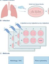
Graphical overview
Background
Cells isolated from animal or human hearts by enzymatic dissociation have provided fundamental insights into the electrophysiology, contractile function, and transcriptional profiles of cardiomyocytes (Liu et al., 2021). However, cells cultured in 2D lack the native environment of the 3D myocardium, which includes intra- and intercellular interactions and syncytial properties that are determinant factors in studying cells, genes, and drug modulations in cardiac pathologies. It is also known that functional and structural preservation of native myocardial tissue ex vivo is dependent on the application of both mechanical preload and electrical stimulation (Brandenburger et al. 2012; Watson et al., 2017; Perbellini et al., 2018; Qiao et al., 2019). Recently, Fischer et al. (2019) established and commercialized a biomimetic ex vivo culture system taking these parameters into account to maintain intact native human myocardial slices in culture for up to four months (Fischer et al., 2019; www.invitrosys.com). This novel technology enables human disease modeling with high temporal and spatial resolution in assessing cellular behavior after pharmacologic or genetic interventions. A drawback of this method is that human tissue is not readily available, with samples generally originating from end-stage heart failure patients with a wide range of comorbidities. Moreover, the patients’ age, genetic background, and medication can be confounding factors during ex vivo analyses. To enable standardized investigational studies, we adapted this system to the culture of non-human primate (NHP) heart slices.
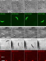
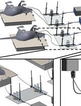
NHP hearts are an excellent surrogate to address basic research questions due to their genetic, metabolic, and physiologic similarities to humans and the ability to control diet, environment, and breeding (Cox et al. 2017). As described in the step-by-step protocol below, left ventricular transmural sections of native NHP hearts are cut into thin tissue slices (300 μm) that are subsequently subjected to physiological preload and electrical stimulation. Under biomimetic culture conditions, a continuous readout of contractile performance and electrophysiologic behavior such as excitability, force-frequency relationship, and effective refractory period can be reached for up to three weeks. Furthermore, as we recently reported, this system can be used as a platform to study critical steps of cardiac regeneration processes in single-cell resolution demonstrating that time- and resource-consuming in vivo studies in animals can be partially bridged (Poch et al. 2022).
Materials and reagents
35 mm Petri dish (Corning Life Sciences, catalog number: CLS430165)
50 mL capped Falcon tube (Corning Life Sciences, catalog number: 352070)
Medium 199, Earle’s salts (M199) (Gibco, catalog number: 11150059)
Insulin-transferrin-selenium (ITS) (Gibco, catalog number: 41400045)
Penicillin-streptomycin (P/S) (Gibco, catalog number: 15140122)
β-Mercaptoethanol (Bio-Rad, catalog number: 1610747)
Histoacryl tissue glue (Braun, catalog number: 04929052)
PET plastic triangles (InVitroSys, Germany)
Isopropyl alcohol (Fischar, catalog number: 08819076)
NaCl (Sigma, catalog number: S5886-1kg)
KCl (Merck, catalog number: 1.04933.0500)
MgCl2·6H2O (AppliChem, catalog number: A1036,0500)
NaH2PO4·H2O (Merck, catalog number: 1.06346.0500)
Glucose·H2O (AppliChem, catalog number: A3730,0500)
CaC12·2H2O (Merck, catalog number: 2382.1000)
BDM (2,3-Butanedione monoxime) (Sigma-Aldrich, catalog number: B0753)
HEPES (AppliChem, catalog number: A 1069,0500)
Filtration unit (Steritop Quick Release, Millipore, S2GPTOSRE)
Agarose low melt (Carl Roth, catalog number: 6351.2)
Cutting buffer (see Recipes)
4% agarose preparation (see Recipes)
Culture medium M199 (see Recipes)
Equipment
Vibratome VT1200S (Leica Biosystems, Germany)
Vibrocheck device (Leica Biosystems, Germany)
MyoDish 1 Tissue Culture System (InVitroSys, Germany)
Incubator (37 °C, 5% CO2) (Thermo Scientific, catalog number: 4110)
Water bath (37 °C)
Razor blade (Wilkinson Sword)
Scalpel (Feather, catalog number: 00636494)
Tweezer (Mmobil®, catalog numbers: ESD-15 and ESD-10)
Software
MyoDish Software (InVitroSys, www.invitrosys.com)
MyoDish Data File Converter v1.1 (inVitroSys, www.invitrosys.com)
LabChart Reader (v8.1.24, www.adinstruments.com)
Microsoft Excel 2019 (www.microsoft.com)
GraphPad Prism 9 (www.graphpad.com)
Procedure
文章信息
版权信息
© 2023 The Author(s); This is an open access article under the CC BY-NC license (https://creativecommons.org/licenses/by-nc/4.0/).
如何引用
Poch, C. M., Dendorfer, A., Laugwitz, K. L. and Moretti, A. (2023). Ex vivo Culture and Contractile Force Measurements of Non-human Primate Heart Slices. Bio-protocol 13(13): e4750. DOI: 10.21769/BioProtoc.4750.
分类
医学 > 心血管疾病 > 心脏组织培养技术
微生物学 > 微生物-宿主相互作用 > 离体模型
药物发现 > 药物筛选 > 抗感染药物
您对这篇实验方法有问题吗?
在此处发布您的问题,我们将邀请本文作者来回答。同时,我们会将您的问题发布到Bio-protocol Exchange,以便寻求社区成员的帮助。