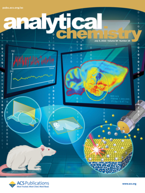
- EN - English
- CN - 中文
Label-free Chemical Characterization of Polarized Immune Cells in vitro and Host Response to Implanted Bio-instructive Polymers in vivo Using 3D OrbiSIMS
利用 3D OrbiSIMS 对体外极化免疫细胞和宿主对体内植入生物结构聚合物的反应进行无标记化学表征
发布: 2023年08月05日第13卷第15期 DOI: 10.21769/BioProtoc.4727 浏览次数: 2078
评审: Aswad KhadilkarNihal Engin VranaVishal Nehru

相关实验方案

研究免疫调控血管功能的新实验方法:小鼠主动脉与T淋巴细胞或巨噬细胞的共培养
Taylor C. Kress [...] Eric J. Belin de Chantemèle
2025年09月05日 3551 阅读

适用于 LC–MS/MS 蛋白质组学分析的大体积细胞培养上清分泌组样品制备方法优化
Basil Baby Mattamana [...] Peter Allen Faull
2025年12月20日 1261 阅读
Abstract
The Three-dimensional OrbiTrap Secondary Ion Mass Spectrometry (3D OrbiSIMS) is a secondary ion mass spectrometry instrument, a combination of a Time of Flight (ToF) instrument with an Orbitrap analyzer. The 3D OrbiSIMS technique is a powerful tool for metabolic profiling in biological samples. This can be achieved at subcellular spatial resolution, high sensitivity, and high mass-resolving power coupled with MS/MS analysis. Characterizing the metabolic signature of macrophage subsets within tissue sections offers great potential to understand the response of the human immune system to implanted biomaterials. Here, we describe a protocol for direct analysis of individual cells after in vitro differentiation of naïve monocytes into M1 and M2 phenotypes using cytokines. As a first step in vivo, we investigate explanted silicon catheter sections as a medical device in a rodent model of foreign body response. Protocols are presented to allow the host response to different immune instructive materials to be compared. The first demonstration of this capability illustrates the great potential of direct cell and tissue section analysis for in situ metabolite profiling to probe functional phenotypes using molecular signatures. Details of the in vitro cell approach, materials, sample preparation, and explant handling are presented, in addition to the data acquisition approaches and the data analysis pipelines required to achieve useful interpretation of these complex spectra. This method is useful for in situ characterization of both in vitro single cells and ex vivo tissue sections. This will aid the understanding of the immune response to medical implants by informing the design of immune-instructive biomaterials with positive interactions. It can also be used to investigate a broad range of other clinically relevant therapeutics and immune dysregulations.
Graphical overview

Background
Mass spectrometry (MS) analysis methods, including liquid chromatography-mass spectrometry (LC–MS) (A. Abuawad et al., 2020), liquid extraction surface analysis mass spectrometry (LESA–MS) (Eikel et al., 2011), matrix-assisted laser desorption/ionization, desorption electrospray ionization, and secondary ion mass spectrometry (SIMS), have been used to detect chemical and biological compounds, such as lipids, amino acids, peptides, and proteins from cells and tissue samples (Rauh, 2012; Cajka and Fiehn, 2014; Passarelli et al., 2017; Kotowska et al., 2020; Meurs et al., 2021). Classes of biomolecules, including proteins, lipids, and metabolites, are vital cellular components that have been characterized and designated to perform specific functions essential to life.
LC–MS-based metabolite analyses typically start with extraction of the metabolites from the biological samples. Analysis of metabolites using LC-MS has been limited because it requires initial liquid extraction procedures and a significant number of cells (1–6 million) to obtain a sufficient signal (Abuawad et al., 2020), leading to a lack of molecular spatial information (Tuli and Ressom, 2009).
The LESA–MS technique is a powerful tool for global, highly sensitive, and multi-analyte analysis ranging from small molecule metabolites to lipids and proteins. This technique has the limitation of involving a solvent-based approach, which allows lipid and small molecule metabolite injection into the analytical instrumentation, as well as poor spatial resolution (~1 mm), rendering single-cell level analysis impossible. For solvent extraction, sample preparation of cells or tissue for metabolomics has a unique protocol for each molecular class. Thus, solvent-extracted metabolites from cells or tissue samples are specific to that extraction protocol (Basu et al., 2018).
SIMS is a direct surface analysis technique that uses a primary ion beam that bombards the surface and generates neutral species and secondary ions (Sigmund, 1969), providing high lateral resolutions of < 100 nm and high surface sensitivity. An electrical field can be used to extract the charge species to obtain mass spectra, using the ion flight times, ion images, and depth profiles in both 2D and 3D (Walker, 2017). SIMS surface analysis has also been established for the quantification of small molecules in biological samples, such as cells and tissue samples.
Time-of-flight secondary ion MS (ToF–SIMS) is a surface analysis technique (Denbigh and Lockyer, 2015; Yoon and Lee, 2018) that provides information-rich mass, depth, and spatial resolution, along with chemical sensitivity. However, ToF–SIMS has insufficient mass accuracy and low mass resolving power for metabolite identification (Green et al., 2011; Shon et al., 2016). To overcome the pitfalls of low mass resolving power and accuracy, the 3D OrbiSIMS technique was developed, which utilizes the SIMS principle with a high mass resolving power (> 240,000 at m/z 200) and accuracy (< 2 ppm), along with the MS/MS capabilities of the OrbiTrapTM mass analyzer (Passarelli et al., 2017).
Recently, the capabilities of the 3D OrbiSIMS instrument as a new means to assess the metabolomic profiles of biological samples have been investigated. For example, Kotowska et al. (2020) used 3D OrbiSIMS imaging and depth profiling to observe a protein monolayer biochip and the depth distribution of proteins in human skin. The platform has also proved its ability to identify metabolite profiling in macrophages treated with different concentrations of the drug amiodarone (Passarelli et al., 2017). Similarly, Suvannapruk et al. (2022) used the novel technique for metabolite identification in individual cells of macrophage subsets. This method of analysis is vital for gaining new insight into metabolomic processes for identifying the metabolites of biological samples.
The development of materials with cell-instructive properties could provide an effective biomaterial-based strategy for modulating cell behavior, to minimize adverse immune responses. Rostam et al. reported that changing the surface topography and chemistry of materials can impact macrophage adhesion and polarization (H. M. Rostam et al., 2015; Hassan M. Rostam et al., 2020). In this paper, we describe for the first time the development of a 3D OrbiSIMS methodology to investigate metabolic changes derived from single anti- and pro-inflammatory macrophages in vitro. We also compare this to metabolic profiles from in vivo anti- and pro-inflammatory macrophages generated at the site of a foreign body response in a mouse model, following exposure to a catheter section coated in known macrophage-instructive surface chemistries. This method aims to directly analyse the metabolic profiles of macrophage phenotypes from in vitro and in vivo studies, with minimal sample preparation steps.
Materials and reagents
Cell culture
T75 flask (Corning 43064, catalog number: 10492371)
50 mL Falcon tube (Sigma-Aldrich, catalog number: T2318)
Pipette tips of various volumes (Fisher Scientific, catalog number: 02-707-401)
Nylon syringe filter 0.22 μm, 25 mm (Minisart®, catalog number: 17845)
Bijou tubes (Thermo Fisher Scientific, catalog number: 129B)
LS columns (Miltenyi Biotech, catalog number: 130-042-401)
Round glass slides (VWR, catalog number: 631-0149)
Glass slides (VWR, catalog number: 631-1553)
24-well plates, uncoated (CytoOne, catalog number: cc77672-7524)
Tissue-Tek Cryomold Moulds (Agar Scientific, catalog number: AGG4580)
Buffy coats from healthy volunteers provided by National Blood Services, Sheffield United Kingdom, after obtaining informed consent and following institutional ethics approval (Research Ethics Committee, Faculty of Medicine and Health Sciences, University of Nottingham; FMHS 425-1221)
Histopaque (Sigma-Aldrich, catalog number: 11191)
Phosphate buffered saline (PBS) (Sigma-Aldrich, catalog number: D8537)
MidiMACSTM separator (Miltenyi Biotech, catalog number: 130-042-302)
MACS® multiStand (Miltenyi Biotech, catalog number: 130-042-303)
CD14 microbeads (Miltenyi Biotech, catalog number: 130-050-201)
Ficoll (Cytiva, catalog number: 1754402)
RPMI 1640 medium (Sigma-Aldrich, catalog number: R0883)
Fetal bovine serum (FBS) (Sigma-Aldrich, catalog number: F9665)
L-glutamine (Sigma-Aldrich, catalog number: G7513)
Penicillin-streptomycin (Sigma-Aldrich, catalog number: P0781)
Interferon gamma, IFN-γ (Bio Techne, catalog number: 285-IF-100)
Granulocyte-macrophage colony-stimulating factor (GM-CSF) (Miltenyi Biotech, catalog number: 130-093-868)
Interleukin 4 (IL-4) (Miltenyi Biotech, catalog number: 130-093-919)
Macrophage colony-stimulating factor (M-CSF) (Miltenyi Biotech, catalog number: 130-096-493)
70% alcohol (any vendor)
Optimal cutting temperature compound (OCT) (Agar Scientific, catalog number: AGR1180)
Copolymer synthesis
Poly(cyclohexyl methacrylate-co-dimethylamino-ethyl methacrylate) (CHMA-DMAEMA), pro-inflammatory macrophage (M1-like)
Poly(cyclohexyl methacrylate-co-isodecyl methacrylate) (CHMA-iDMA), anti-inflammatory macrophage (M2-like)
Liquid nitrogen
Ammonium formate (Sigma-Aldrich, catalog number: 70221)
150 mM Ammonium formate (see Recipes)
MACS buffer (see Recipes)
Animal study
Clinical-grade silicon catheter 13 mm diameter (Teleflex Medical, catalog number: RUSCH170003)
Polymer synthesis
Dichloromethane (any vendor)
MED1-161 SILICONE PRIMER (Nusil)
Female BALB/c strain 19–22 g mice (Charles River)
Equipment
Stripette (Greiner Bio-One, catalog number: 760160)
Stripette gun (any vendor)
Micropipette (any vendor)
Forceps (any vendor)
Cell culture hood, class II (any vendor)
Scissors (any vendor)
Water bath (any vendor)
Centrifuge (any vender)
Automated cell counter (any vendor)
Cell culture incubator (SANYO, model: MC0-18A1C)
UV Clave (any vendor)
Refrigerator (4 °C) (any vendor)
Freezer (-80 °C) (SANYO, model: MDF-C8V1)
Cryostat CM3050 (Leica Microsystems)
Freeze dryer (any vendor)
Dip coating unit (Holmarc, model: HO-TH-01)
Vacuum oven (Thermo Fisher Scientific)
3D OrbiSIMS (IONTOF GmbH, Germany and Thermo Fisher Scientific, Germany)
Software
SurfaceLab software version 7.1 (ION-TOF, Germany), which utilizes the Thermo Fisher provided application programming interface
LIPID MAPS software (https://www.lipidmaps.org)
Procedure
文章信息
版权信息
© 2023 The Author(s); This is an open access article under the CC BY-NC license (https://creativecommons.org/licenses/by-nc/4.0/).
如何引用
Suvannapruk, W., Edney, M. K., Fisher, L. E., Luckett, J. C., Kim, D. H., Scurr, D. J., Ghaemmaghami, A. M. and Alexander, M. R. (2023). Label-free Chemical Characterization of Polarized Immune Cells in vitro and Host Response to Implanted Bio-instructive Polymers in vivo Using 3D OrbiSIMS. Bio-protocol 13(15): e4727. DOI: 10.21769/BioProtoc.4727.
分类
免疫学 > 免疫细胞功能 > 巨噬细胞
生物科学 > 生物技术 > 质谱
您对这篇实验方法有问题吗?
在此处发布您的问题,我们将邀请本文作者来回答。同时,我们会将您的问题发布到Bio-protocol Exchange,以便寻求社区成员的帮助。
Share
Bluesky
X
Copy link


