

Systematic Analysis of Smooth Muscle and Cartilage Ring Formation during Mouse Tracheal Tubulogenesis
小鼠气管插管过程中平滑肌和软骨环形成的系统分析
(*contributed equally to this work) 发布: 2023年07月05日第13卷第13期 DOI: 10.21769/BioProtoc.4711 浏览次数: 1537
评审: Anonymous reviewer(s)
参见作者原研究论文
The authors used this protocol in:

Jul 2018


Abstract
The trachea tube is the exclusive route to allow gas exchange between the external environment and the lungs. Recent studies have shown the critical role of mesenchymal cells in tracheal tubulogenesis. Improved methods for studying the dynamics of the tracheal mesenchyme development are needed to investigate the cellular and molecular mechanisms during tracheal tubulogenesis. Here, we describe a detailed protocol for a systematic analysis of tracheal tube development to enable observing tracheal smooth muscle (SM) and cartilage ring formation. We describe immunostaining, confocal and stereomicroscopy imaging, and quantitative methods to study the process of tracheal SM and cartilage ring development, including SM cell alignment, polarization, and changes in cell shape as well as mesenchymal condensation. The technologies and approaches described here not only improve analysis of the patterning of the developing trachea but also help uncover the mechanisms underlying airway disease. This protocol also provides a useful technique to analyze cell organization, polarity, and nuclear shape in other organ systems.
Keywords: Trachea (气管)Background
Understanding tracheal formation is a fundamental goal in the field of pulmonary development and disease. The trachea consists of mesoderm-derived smooth muscle (SM), cartilage, and connective tissue, as well as endoderm-derived epithelium (Brand-Saberi and Schafer, 2014). SM is positioned dorsally to control tracheal contraction, whereas the cartilage rings are located ventrally to prevent airway collapse (Hines et al., 2013; Yin et al., 2018 and 2019).
In humans, defects in the formation of the tracheal tube have been reported to lead to tracheomalacia, tracheostenosis, or complete tracheal ring deformity, which are characterized by a deficiency of the supporting cartilage or narrowing of the tracheal lumen and may lead to respiratory distress and death (Landing and Dixon, 1979; Fraga et al., 2016; Sinner et al., 2019).
In mice, tracheal SM differentiation starts by embryonic day 11.5 (E11.5) with the appearance of α-smooth muscle actin (αSMA) positive cells at the dorsal side (Hines et al., 2013; Yin et al., 2018). Newly differentiated tracheal SM cells exhibit round shapes and are not well organized (Yin et al., 2018 and 2019). They progressively develop into spindle-shaped cells that circumferentially align the tube (Yin et al., 2018 and 2019). During tracheal elongation, differentiated SM cells proliferate with increased area of SM stripes from E11.5 to P0 (Yin et al., 2018).
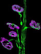
Tracheal cartilage development initiates as early as E9 (Elluru and Whitsett, 2004). Uncondensed SOX9+ mesenchymal cells appear and are restricted to the ventral trachea as early as E10.5 (Hines et al., 2013). Both Sox9 mRNA levels and the number of SOX9+ mesenchymal cells increase at E12.5 (Hines et al., 2013). By E13.5, SOX9+ mesenchymal cells condense to resemble cartilaginous rings with the number of 11–13. The number of C-shaped rings appears to be unchanged, whereas both the distance between rings and ring width increase during tracheal elongation (Yin et al., 2019). By E15.5, SOX9+ mesenchymal cells start differentiating into chondrocytes characterized by positive alcian blue staining, as well as aggrecan and type II collagen expression (Park et al., 2010; Yin et al., 2019).
Analysis of tracheal formation has mostly focused on cell condensation, differentiation, proliferation, and apoptosis by using tissue sections (Park et al., 2010; Hines et al., 2013; Lin et al., 2014). These studies have provided information on cell behavior in two-dimensional views, allowing for a better understanding of the cellular and molecular mechanisms underlying tracheal tubulogenesis. The method we present here for whole-mount immunostaining and imaging overcomes some of their limitations; for example, the precise sites of the three-dimensional trachea are not properly located. Thus, it is presently unclear how cells are organized in the whole trachea. Another not fully investigated question is the characteristics of SM cells. Although SM cell differentiation and proliferation have been tested (Hines et al., 2013; Gerhardt et al., 2018), cell alignment, polarization, and changes in cell shape in tracheal development remain largely unknown. To analyze tissue structure and cell organization, we provide a detailed protocol for examining SM cell organization and changes in cell shape as well as mesenchymal cell condensation, an important process during tracheal cartilage formation (Yin et al., 2019).
Materials and reagents
Coverslips (Thermo Fisher Scientific, catalog number: 11961988)
Adhesion slides (Thermo Fisher Scientific, catalog number: 10219280)
10 cm Petri dish (Corning, catalog number: 430167)
Pipette (Sigma-Aldrich, catalog number: Z331759-1PAK)
Cy3-conjugated anti-mouse αSMA (Sigma-Aldrich, catalog number: C6198)
Anti-rat CDH1 (E-cadherin) (Santa Cruz, catalog number: sc-59778)
Anti-rabbit SOX9 (SRY-box transcription factor 9) (Millipore, catalog number: AB5535)
Sheep anti-GM130/GOLGA2 (Golgin subfamily A member 2) (R&D systems, catalog number: AF8199)
Paraformaldehyde (PFA) (Sigma-Aldrich, catalog number: 30525-89-4)
Dimethyl sulfoxide (DMSO) (Sigma-Aldrich, catalog number: D8418)
Mowiol (Millipore, catalog number: 475904)
30% hydrogen peroxide (H2O2) (Sigma-Aldrich, catalog number: 7722-84-1)
Methanol (Sigma-Aldrich, catalog number: 34860)
Bovine serum albumin (BSA) (Thermo Fisher Scientific, catalog number: 30036578)
Fetal bovine serum (FBS) (Gibco, catalog number: 10270-106)
4',6-Diamidino-2-Phenylindole, dilactate (DAPI) (Thermo Fisher Scientific, catalog number: D3571)
Phosphate buffered saline (PBS) (Capricorn Scientific, catalog number: PBS-1A)
Hanks’ balanced salt solution (HBSS) (Sigma-Aldrich, catalog number: H6648)
Triton X-100 (Sigma-Aldrich, catalog number: 9036-19-5)
Glycerol (Sigma-Aldrich, catalog number: USA56-81-5)
Benzyl benzoate (Sigma-Aldrich, catalog number: B6630)
Benzyl alcohol (Sigma-Aldrich, catalog number: 305197)
Benzyl benzoate (Sigma-Aldrich, catalog number: B6630-1L)
Sucrose (Sigma-Aldrich, catalog number: 57-50-1)
Tissue-Tek OCT (SAKURA, catalog number: 4583)
Goat anti-rabbit IgG (H+L) highly cross-adsorbed secondary antibody, Alexa FluorTM 488 (Thermo Fisher Scientific, catalog number: A-11034)
Donkey anti-sheep IgG (H+L) secondary antibody, Alexa FluorTM 647 (Thermo Fisher Scientific, catalog number: A-21448)
Goat anti-rat IgG (H+L) secondary antibody, Alexa FluorTM 568 (Thermo Fisher Scientific, catalog number: A-11077)
Phosphate buffered saline (PBS) (see Recipes)
4% formaldehyde solution (see Recipes)
Permeabilization solution (see Recipes)
10% sucrose (see Recipes)
30% sucrose (see Recipes)
Mowiol mounting medium (see Recipes)
0.3% Triton X-100/PBS solution (see Recipes)
5% FBS/PBS/0.5% Triton X-100/3% BSA solution (see Recipes)
70% ethanol solution (see Recipes)
DMSO:methanol (1:4) solution (see Recipes)
H2O2/DMSO/methanol (1:1:4) solution (see Recipes)
80% methanol solution (see Recipes)
50% methanol solution (see Recipes)
Benzyl alcohol:benzyl benzoate (BABB) (1:2) solution (see Recipes)
5% FBS/PBS/3% BSA blocking solution (see Recipes)
Equipment
Scissors (FST, catalog number: 15011-12)
Forceps (FST, catalog number: 11252-20)
Stereoscopic dissecting microscope (Zeiss, model: Stemi 305)
Stereomicroscope (Nikon, model: SMZ25)
Upright laser scanning confocal microscope (Zeiss, model: LSM 880) or cryostat (Leica, model: CM1950)
Quantification of immunofluorescence intensity of lumen area, tube length, SM area, SM cell orientation, nuclear, aspect ratio (NAR), and Golgi-apparatus position relative to the nucleus was performed using ImageJ (Version 1.8.0.172) (Schneider et al., 2012)
Software
ImageJ, GraphPad Prism (Version 8.0.2)
Zeiss ZEN (Version 2.3)
Procedure
文章信息
版权信息
© 2023 The Author(s); This is an open access article under the CC BY-NC license (https://creativecommons.org/licenses/by-nc/4.0/).
如何引用
Wu, H., Wang, P., Liu, Z., Lu, C. and Yin, W. (2023). Systematic Analysis of Smooth Muscle and Cartilage Ring Formation during Mouse Tracheal Tubulogenesis. Bio-protocol 13(13): e4711. DOI: 10.21769/BioProtoc.4711.
分类
发育生物学 > 形态建成 > 器官形成
细胞生物学 > 组织分析 > 组织染色
细胞生物学 > 细胞成像 > 共聚焦显微镜
您对这篇实验方法有问题吗?
在此处发布您的问题,我们将邀请本文作者来回答。同时,我们会将您的问题发布到Bio-protocol Exchange,以便寻求社区成员的帮助。