

Cycloheximide (CHX) Chase Assay to Examine Protein Half-life
环己酰亚胺(CHX)追踪测定法检测蛋白质半衰期
发布: 2023年06月05日第13卷第11期 DOI: 10.21769/BioProtoc.4690 浏览次数: 16666
评审: Ralph Thomas BoettcherDeepali BhandariAnonymous reviewer(s)
参见作者原研究论文
The authors used this protocol in:
Oct 2022


Abstract
Cycloheximide (CHX) is a small molecule derived from Streptomyces griseus that acts as fungicide. As a ribosome inhibitor, CHX can restrict the translation elongation of eukaryotic protein synthesis. Once protein synthesis is inhibited by CHX, the level of intracellular proteins decreases by degradation through the proteasome or lysosome system. Thus, the CHX chase assay is widely recognized and used to observe intracellular protein degradation and to determine the half-life of a given protein in eukaryotes. Here, we present a complete experimental procedure of the CHX chase assay.
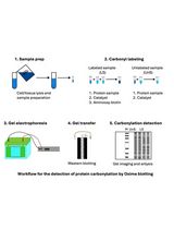
Graphical overview

Background
Regulation of protein abundance is crucial for the maintenance of normal cell function; all proteins in the cell are constantly being degraded and replaced. Specifically, rapid protein degradation plays an important role in regulating protein abundance in a variety of cellular processes, including cell cycle, signal transduction, and differentiation (Goldberg, 2003). Studies on protein degradation rate and half-life are important for understanding protein function, developing drugs to regulate the degradation of disease-specific proteins, and researching small molecule compounds to regulate protein levels (Paiva and Crews, 2019). There are two main protein degradation pathways in eukaryotic cells—the ubiquitin-proteasome pathway and the autophagy-lysosome pathway. The former is the main pathway of protein degradation in cells and is involved in the degradation of various proteins (Glickman and Ciechanover, 2002). It degrades proteins in cells in a specific, highly complex mechanism, and can be reversed by deubiquitinating enzymes (Sun et al., 2020). Autophagy-lysosome pathway is a self-degradation process, which degrades proteins through lysosomal mediation, by coating them and forming an autophagic lysosome, to fulfill the metabolic needs of the cells. Both pathways play vital roles in maintaining the normal protein metabolism of cells, and abnormal protein degradation may lead to many diseases, especially neurodegenerative disorders and cancer (Huang et al., 2018; Park et al., 2020).
The half-life of proteins varies widely, from a few minutes to days, and different rates of protein degradation are important for protein function. The classical method for protein degradation detection is the pulse-chase assay (Fritzsche and Springer, 2014); this assay is based on the addition of radionuclide-labeled amino acids, such as 35S-labeled methionine, to cell culture medium. The change process of a specific protein in the cells can be detected by immunoblotting or other methods. However, this method uses isotopes, which have some negative effects on the health and safety of researchers, and the operation process is complicated. Another common approach to study protein degradation is the protein translation blocker cycloheximide (CHX), which we will describe in detail later. In recent years, mass spectrometry technology has developed to detect the degradation of the primary structure of proteins. Therefore, protein degradation detection by mass spectrometry can be used to study protein structure, function, and quality control (Spradlin et al., 2021). In addition, it has been reported that CuAAC (copper-catalyzed azide–alkyne cycloadditions) can be used to label newly synthesized proteins in cells and to study protein degradation (Lu et al., 2013). Besides, protein degradation can be studied through the addition of pharmacological inhibitors of the degradation pathway to cells (Hanzl and Winter, 2020). For example, proteasome inhibitors such as MG-132, epoxomicin, and bortezomib block protein degradation via the ubiquitin-proteasome pathway. Inhibitors such as chloroquine and bafilomycin A1 work by neutralizing the acidic pH in the lysosome, which is necessary for the function of lysosomal protease.
Here, we describe a method that inhibits protein synthesis by adding CHX to assess the degradation of the target protein over time. CHX is a global translational inhibitor that can block the eukaryotic ribosome translocation process (Schneider-Poetsch et al., 2010). CHX is a product of Streptomyces griseus that can be used to determine the half-life of proteins or enzymes in molecular biology. It is noteworthy that CHX can inhibit eukaryotic protein synthesis but not prokaryotic protein synthesis. The advantages of CHX are that it is easy to operate, does not require considering the effect of ongoing protein synthesis, and allows for high-throughput screening. On the other hand, its main disadvantages are i) blocking all protein translation without specificity, and ii) having high cytotoxicity, able to cause large-scale cell death after prolonged treatment, so it is not suitable for proteins with a slow degradation rate. Therefore, CHX needs to be selected according to the appropriate conditions.
CHX chase assay visualizes protein degradation kinetics through CHX treatment over different times and western blotting analysis. Briefly, cells are treated with CHX at a suitable concentration, and then harvested at different timepoints. Next, western blot is employed to analyze the expression levels of a given protein. A short half-life protein will decrease in abundance over time, while a long half-life protein could exhibit little change in abundance. Thus, the CHX chase assay visualizes protein degradation efficiently without the use of radioactive isotopes, which provides an easy and reliable means to observe the change of intracellular protein levels and therefore could contribute to the study of therapeutic targets for a range of diseases.
Materials and reagents
Materials
Cell culture dish, 100 mm diameter (NEST, catalog number: 704001)
12-well plate (NEST, catalog number: 712011)
1,000 μL blue tip (Biosharp, catalog number: BS-1000-T)
200 μL yellow tip (Biosharp, catalog number: BS-200-T)
10 μL clear tip (Biosharp, catalog number: BS-10-T)
1.5 mL tube (AXYGEN, catalog number: AXYMCT150CS)
0.6 mL tube (AXYGEN, catalog number: AXYMCT060C)
Medical X-ray film (FUJIFILM, catalog number: SUPER RX-N)
Cell scraper (BIOLGIX, catalog number: 70-1180)
Cells
A549 cells (American Type Culture Collection)
Antibodies and plasmids
Anti-Tyk2 antibody (Cell Signaling Technology, catalog number: 14193; 1:1,000)
Anti-AHI1 antibody (Santa Cruz, catalog number: sc-515382; 1:500)
Anti-tubulin antibody (Proteintech, catalog number: 66031-1-Ig; 1:3,000)
Goat anti-rabbit IgG (H+L) HRP (Bioworld, catalog number: BS13278; 1:10,000)
Goat anti-mouse IgG (H+L) HRP (Bioworld, catalog number: BS12478; 1:10,000)
shCtrl plasmid (RNAi-Ready pSIREN-RetroQ-ZsGreen vector)
shAHI1 plasmid
shAHI1(#1)-forward: 5'-CACCGCGGAGACATTATCCGAGTGTTCGAAAACACTCGGATAATGTCTCCG-3'
shAHI1(#1)-reverse: 5'-AAAACGGAGACATTATCCGAGTGTTTTCGAACACTCGGATAATGTCTCCGC-3'
shAHI1(#2)-forward: 5'-CACCGCCATATTGGTCCGACAGTTTCGAAAAACTGTCGGACCAATATGGC-3'
shAHI1(#2)-reverse: 5'-AAAAGCCATATTGGTCCGACAGTTTTTCGAAACTGTCGGACCAATATGGC-3'
Reagents
Dulbecco’s modified Eagle medium (DMEM basic medium) (Gibco, catalog number: C11995500BT)
Fetal bovine serum (FBS) (PAN, catalog number: P30-3302)
Penicillin-streptomycin (Biosharp, catalog number: BL505A)
0.25% trypsin-EDTA (Biosharp, catalog number: BL512A)
Cycloheximide (CHX) (Sigma, catalog number: C7698-1G)
SuperSignal West Dura Extended kit (Thermo Scientific)
LongTrans (UCallM, catalog number: TF07)
Dimethyl sulfoxide for cell culture (DMSO) (panreac applichem, catalog number: 67-68-5)
Bradford (Sangon Biotech, catalog number: C900164-0200)
Non-fat milk (Biosharp, catalog number: BS102-500g)
BSA (Solarbio, catalog number: A8020)
Serum-free cell freezing medium (NCM Biotech, catalog number: C40100)
KCl (Sangon Biotech, catalog number: 7447-40-7)
NaCl (Sangon Biotech, catalog number: 7647-14-5)
EDTA (Solarbio, catalog number: 9002-07-7)
NP40 (Solarbio, catalog number: 9016-45-9)
Inhibitor cocktail (Sigma, catalog number: 58914000)
NaF (Sangon Biotech, catalog number: 7681-49-4)
PMSF (Solarbio, catalog number: 329-98-6)
Acr-Bis (30%) (Biosharp, catalog number: BL513B)
TEMED (Beyotime, catalog number: SY728)
APS (Sangon Biotech, catalog number: 7727-54-0)
SDS (Solarbio, catalog number: 151-21-3)
Glycine (Solarbio, catalog number: 56-40-6)
Tris base (Solarbio, catalog number: 77-86-1)
Tween 20 (Sangon Biotech, catalog number: 9005-64-5)
β-mercaptoethanol (Sigma, catalog number: 60-24-2)
Isopropyl alcohol (Sangon, catalog number: A507048-0500)
PageRuler prestained protein ladder (Thermo Fisher Scientific, catalog number: 26616)
Na3VO4 (Sangon Biotech, catalog number: 13721-39-6)
Na2HPO4 (Solarbio, catalog number: 7558-79-4)
KH2PO4 (Solarbio, catalog number: 7778-77-0)
Bromophenol blue (Solarbio, catalog number: 34725-61-6)
Glycerol (Solarbio, catalog number: G8190-500 ml)
HCl (Yonghua, catalog number: 210101204)
10× PBS Buffer (see Recipes)
Lysis buffer (1% NP40) (see Recipes)
8% separation gel (see Recipes)
5% stacking gel (see Recipes)
10× running buffer (see Recipes)
10× transfer buffer (see Recipes)
100 mM PMSF (see Recipes)
10% APS (see Recipes)
Stacking mix (see Recipes)
3× loading buffer (see Recipes)
SDS-sample buffer (see Recipes)
1× PBST buffer (see Recipes)
1% NP40 washing buffer (see Recipes)
1 M Tris-HCl (see Recipes)
Equipment
37 °C, 5% CO2 forced-air incubator (Thermo Scientific, catalog number: 3111)
Electric thermostatic water tank (Shanghai Jing Hong, catalog number: DK-8D)
Dry thermostat (AOSHENG, catalog number: MK-10)
SDS-PAGE gel casting apparatus (Tanon, catalog number: VE-180)
Protein electrophoresis and blotting instruments (Tanon, catalog number: EPS 300)
Centrifuge (Eppendorf, catalog number: 5425R)
Software
Microsoft PowerPoint
ImageJ
GraphPad Prism
Procedure
文章信息
版权信息
© 2023 The Author(s); This is an open access article under the CC BY-NC license (https://creativecommons.org/licenses/by-nc/4.0/).
如何引用
Readers should cite both the Bio-protocol article and the original research article where this protocol was used:
- Miao, Y., Du, Q., Zhang, H. G., Yuan, Y., Zuo, Y. and Zheng, H. (2023). Cycloheximide (CHX) Chase Assay to Examine Protein Half-life. Bio-protocol 13(11): e4690. DOI: 10.21769/BioProtoc.4690.
- Zhang, H. G., Wang, B., Yang, Y., Liu, X., Wang, J., Xin, N., Li, S., Miao, Y., Wu, Q., Guo, T., et al. (2022). Depression compromises antiviral innate immunity via the AVP-AHI1-Tyk2 axis. Cell Res 32(10): 897-913.
分类
细胞生物学 > 细胞新陈代谢 > 其它化合物
生物化学 > 蛋白质 > 降解
您对这篇实验方法有问题吗?
在此处发布您的问题,我们将邀请本文作者来回答。同时,我们会将您的问题发布到Bio-protocol Exchange,以便寻求社区成员的帮助。