

Modified Pseudo-Schiff Propidium Iodide for Staining the Shoot Apical Meristem in Arabidopsis
改良的拟Schiff碘化丙啶对拟南芥茎尖分生组织的染色
发布: 2023年05月05日第13卷第9期 DOI: 10.21769/BioProtoc.4672 浏览次数: 2273
评审: Ansul LokdarshiRicardo Urquidi CamachoPablo Bolanos-Villegas
参见作者原研究论文
The authors used this protocol in:
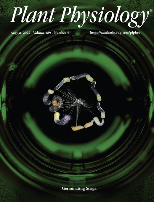
Aug 2022


Abstract
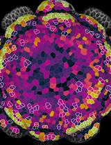
Visualization of cell structure with fluorescent dye for characterizing cell size, shape, and arrangement is a common method to study tissue morphology and morphogenesis. In order to observe shoot apical meristem (SAM) in Arabidopsis thaliana by laser scanning confocal microscopy, we modified the pseudo-Schiff propidium iodide staining method by adding a series solution treatment to stain the deep cells. The advantage of this method is mainly reflected by the direct observation of the clearly bounded cell arrangement and the typical three-layer cells in SAM without the traditional tissue slicing.
Keywords: Shoot apical meristem (SAM) (茎尖分生组织(SAM))Background
Most of the above-ground plant organs are derived from the shoot apical meristem (SAM) (Steeves and Sussex, 1989; Dinneny and Benfey, 2008). As a key tissue involved in plant growth and development, it plays a role in maintaining the ability of stem cells to divide continuously (Fletcher, 2018; Hu et al., 2018), controlling the initiation of organs development (Spinelli et al., 2011; Balkunde et al., 2017), accurately regulating the spatio-temporal specific gene expression (Mayer et al., 1998; Long et al., 1996; Williams, 2021), and integrating the signals from various cell types (Zhao et al., 2010; Azizi et al., 2015; Chung et al., 2019). Thus, exploring the mechanism of complex regulation patterns in the meristem is inseparable from observation of meristem at the cellular level.
Unlike the inflorescence meristem, the SAM of Arabidopsis is tightly wrapped by the surrounding leaf primordia and leaves, which makes it difficult to observe its cellular structure in its deeper part (Moreno et al., 2006). Scientists usually observe the cell structure and morphogenesis of SAM by semithin sections or tissue clearing, but only one cell layer or the surface layer of the SAM is observed (Koi and Kato, 2007; Zhang et al., 2021; Wang et al., 2022). To visualize three-dimensional structure of plant tissues, scientists prefer to use fluorescent dyes, such as propidium iodide, to stain tissues for optical section for laser scanning confocal microscopy (Haseloff, 2003; Truernit et al., 2008; Prunet et al., 2016; Shi et al., 2016).
Previously, we observed the cell structure of SAM and inflorescence meristem in Arabidopsis by modifying the pseudo-Schiff propidium iodide staining method (Li et al., 2022). Compared with traditional methods, this method presents several advantages. First, it allows the observation of the different cell layers in the whole SAM, which is a continuous and complete structure. Second, the cellular structure of the initial leaf primordium is observable except for the SAM. Third, unlike the traditional method that takes seven days, this method is simple in terms of procedure and time saving, taking only three days to complete. It can be foreseen that this method will likely be applicable to the observation of other tissues or SAM of other plant species. The operation details are described in the following section.
Materials and Reagents
1.5 mL centrifuge tubes (Chenglin, catalog number: CL22091809)
Microscope slides (CITOTEST, catalog number: 10127101P-G)
Microscope cover glass (CITOTEST, catalog number: 10212222C)
Qualitative filter paper (Beimu, catalog number: 101)
60 mm round culture dishes (Jindian, catalog number: 16021-1)
Aluminum foil for experiment (Zaowufang, catalog number: 615)
1 mL syringe (Zhi Yu, catalog number: V500111)
Single edge blades (Personna, catalog number: 84-0701)
Sodium hypochlorite solution (Aladdin, catalog number: S101636)
Murashige and Skoog basal medium (Sigma, catalog number: M5519)
Sucrose (Hushi, catalog number: 10021418)
Potassium hydroxide (Hushi, catalog number: 10017008)
Methanol (Aladdin, catalog number: M116115)
Acetate (Aladdin, catalog number: A116172)
Ethanol (Aladdin, catalog number: E130059)
Hydrochloric acid (Aladdin, catalog number: H399657)
Sodium hydrogen bisulfite (Hushi, catalog number: 10019018)
Phytagel (Sigma, catalog number: P8169)
Periodic acid (Sigma, catalog number: P0430)
Propidium iodide (Sigma-Aldrich, catalog number: P4170)
Chloral hydrate C-IVN (Sigma-Aldrich, catalog number: 15307)
Glycerol (Sigma-Aldrich, catalog number: G7893)
Gum arabic from acacia tree (Sigma, catalog number: G9752)
Solid MS medium (see Recipes)
Fixative solution (see Recipes)
1% periodic acid solution (see Recipes)
Pseudo-Schiff propidium iodide solution (see Recipes)
Chloral hydrate solution (see Recipes)
Hoyer′s solution (see Recipes)
Ethanol solutions (see Recipes)
Equipment
Tweezers (Venus, #5)
Dissecting microscope (Zeiss, model: Axio Zoom V16)
Laser scanning confocal microscope (Zeiss, model: 780)
Growth chamber (Percival, model: CU36L5)
Fume hood
Clean bench
Procedure
文章信息
版权信息
© 2023 The Author(s); This is an open access article under the CC BY-NC license (https://creativecommons.org/licenses/by-nc/4.0/).
如何引用
Li, R., Li, Q. and Ma, L. (2023). Modified Pseudo-Schiff Propidium Iodide for Staining the Shoot Apical Meristem in Arabidopsis. Bio-protocol 13(9): e4672. DOI: 10.21769/BioProtoc.4672.
分类
植物科学 > 植物发育生物学 > 形态建成
发育生物学 > 形态建成 > 细胞结构
细胞生物学 > 细胞结构 > 细胞核
您对这篇实验方法有问题吗?
在此处发布您的问题,我们将邀请本文作者来回答。同时,我们会将您的问题发布到Bio-protocol Exchange,以便寻求社区成员的帮助。