

Long-term in toto Imaging of Cellular Behavior during Nerve Injury and Regeneration
神经损伤和再生过程中细胞行为的长期整体成像
发布: 2023年05月05日第13卷第9期 DOI: 10.21769/BioProtoc.4665 浏览次数: 2412
评审: Nafisa M. JadavjiXiaochen SunWenyang Li
参见作者原研究论文
The authors used this protocol in:

Mar 2021
Advertisement


Abstract
Accidental wounding of the peripheral nervous system leads to acute neural dysfunction. Normally, chronic deficits are overcome because peripheral nerves naturally regenerate. However, various genetic and metabolic defects can impair their natural regenerative capacity, which may be due to neuron-extrinsic mechanisms. Therefore, characterizing the behavior of multiple cells during nerve injury and repair in vivo is a pressing need in regenerative medicine. Here, we detail a method for precise wounding of sensory axons in zebrafish, followed by high-resolution in toto long-term quantitative videomicroscopy of neurons, Schwann cells, and macrophages. This protocol can be easily adapted to study the effects of targeted genetic or metabolic disruptions in zebrafish and other suitable organisms, as well as for screening pharmacological agents with therapeutic potential.
Graphical overview

Background
The peripheral nervous system communicates sensory cues to the brain. It therefore mediates organismal responses to changes in the body’s internal and external environments (Handler and Ginty, 2021; Elias and Abdus-Saboor, 2022; Prescott and Liberles, 2022). Sensory neurons must maintain functionality throughout the life of the organism despite stress and trauma. Although acute loss of integrity of these neurons occurs persistently, animals normally overcome chronic neurological dysfunctions via effective repair (Johnson et al., 2005). By contrast, repair is normally extremely limited in the central nervous system (Varadarajan et al., 2022). Peripheral nerve wounding triggers dynamic changes in the behavior of Schwann cells as well as the recruitment of other cells including macrophages, which together resolve injury, initiate repair, and enhance regeneration (Abdo et al., 2019). In recent studies, we have combined genetic and chemical perturbations, transgenic markers, and high-resolution live imaging to characterize nerve injury and regeneration in larval zebrafish (Lozano-Ortega et al., 2018; Tian and López-Schier, 2020; Xiao et al., 2015a). Furthermore, we discovered that Schwann cells are important but not essential for sensory nerve regeneration (Tian et al., 2020b; Xiao et al., 2015b). Moreover, we revealed fast recruitment of macrophages to the wound (Tian et al., 2020). We have also discovered that blocking the degradation of axons severed from the neuronal cell body has no significant impact on nerve repair (Tian et al., 2020). Our finding that loss of the pro-degenerative protein Sarm1 accelerates axonal regeneration may have important implications for the development of therapies aimed at improving neural repair in humans.
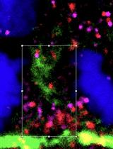
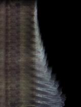
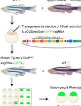
The zebrafish larva is a powerful vertebrate experimental system for studies of neuronal wounding and regeneration (Rieger and Sagasti, 2011; Cardozo et al., 2017; Cigliola et al., 2020; González and Allende, 2021). Spatial and temporal control of injury can be done by laser-mediated axon transection or neuronal ablation. Axonal injury triggers fast degeneration of the part of the axon detached from the cell body, followed by regenerative growth of the proximal stump. In several previous studies, we used the mechanosensory lateral line in larval zebrafish to perform nerve wounding using an ultraviolet laser, and live microscopy in transgenic animals expressing fluorescent markers to track relevant cellular populations during injury resolution and repair (Figures 1 and 2). Of note, laser-mediated axon severing is a powerful technique but it suffers from the fact that both proximal and distal axon fragments are immediately cauterized. This is unlikely to occur during physical injury, when axoplasmic spillage may trigger different responses from the Schwann cells situated in proximity to the damaged nerve. Nevertheless, the precise spatiotemporal control of injury and simultaneous live imaging that this protocol allows vastly overcome this potential shortcoming. Therefore, we present a detailed protocol for facile implementation of laser-mediated microsurgery of sensory axons and in toto imaging of subsequent responses by axons, glia, and macrophages (Figure 3 and 4, and Videos 1 and 2). This protocol uses 5-day-post-fertilization (5 dpf) zebrafish Danio, but it can be equally used in older animals and other specimens of similar characteristics, including Oryzias, Astyanax, or Danionella.
An ultraviolet laser is directed to the target through a high numerical aperture objective lens mounted on a spinning-disk confocal microscope. However, a point-scanning single- or two-photon confocal microscope will be useful. Targeting is guided by the fluorescence from stable expression of EGFP or RFP using a lateralis afferent neuron-specific enhancer. However, mosaic expression of a fluorescent protein using pan-neuronal genetic control elements, for instance the widely employed NeuroD, Ngn1, or HuC, is also suitable.
Materials and Reagents
Cover glass–bottomed mini dish (MatTek, catalog number: p35G-1.0-14-c)
Petri dishes (Greiner Bio-One, catalog number: G180535A/01889)
Plastic strainer (ZM Systems, Product code: teastrain15)
Plastic Pasteur pipettes
Tricaine (MS-222) (25× stock solution) (PharmQ, UK)
Low-melting agarose (1% M/V) (Sigma-Aldrich, catalog number: A9414-100G)
Sodium chloride (NaCl) (Merck KGaA, catalog number: 1.06404.5000)
Potassium chloride (KCl) (Sigma-Aldrich, P9541-500G)
Calcium chloride dihydrate (CaCl2·2H2O) (Sigma-Aldrich, catalog number: C3306-250G)
Magnesium sulfate (MgSO4) (Sigma-Aldrich, catalog number: M5921 500G)
Trizma (Tris base) (Sigma-Aldrich, catalog number: T1503-1KG)
HCl (Sigma-Aldrich, catalog number: H1758-500ML)
Plasmids UAS:EGFP, SILL:mCherry; mfap4:EGFP
Fish lines: Casper, Tg[UAS:EGFP], Tg[SILL:mCherry], Tg[gSAGFF202A], Tg[ mfap4:EGFP]
E3 embryo medium (10×) (see Recipes)
Tris-HCl (1 M pH 9.0) (see Recipes)
Tricaine (MS-222) (25×) (see Recipes)
Low-melting agarose (1% w/v) (see Recipes)
Equipment
Incubator set at 28.5 °C
Fine forceps
Optically-clear polystyrene 90 mm Petri dishes
Plastic transfer pipettes
Microwave oven
100 mL glass bottle
Handmade 1-eyelash brush
Microinjection system
Microscopes:
Upright stereomicroscope (e.g., ZEISS Stemi SV 11) equipped with fluorescent-light source and a filter set for GFP and RFP/Cherry.
Spinning-disk microscope confocal microscope (ZEISS) with an iLasPulse laser system (Roper Scientific SAS) and a temperature-control system.
Procedure
文章信息
版权信息
© 2023 The Authors; exclusive licensee Bio-protocol LLC.
如何引用
Readers should cite both the Bio-protocol article and the original research article where this protocol was used:
- Tian, W., González-Suarez, A. and López-Schier, H. (2023). Long-term in toto Imaging of Cellular Behavior during Nerve Injury and Regeneration. Bio-protocol 13(9): e4665. DOI: 10.21769/BioProtoc.4665.
- Asgharsharghi, A., Tian, W., Haehnel-Taguchi, M. and López-Schier, H. (2021). Sarm1 is dispensable for mechanosensory-motor transformations in zebrafish. MicroPubl Biol. DOI: 10.17912/micropub.biology.000369
分类
神经科学 > 细胞机理
发育生物学 > 细胞生长和命运决定 > 再生
细胞生物学 > 细胞成像 > 荧光
您对这篇实验方法有问题吗?
在此处发布您的问题,我们将邀请本文作者来回答。同时,我们会将您的问题发布到Bio-protocol Exchange,以便寻求社区成员的帮助。