

Analysis of Mouse Brain Sections by Live-cell Time-lapse Confocal Microscopy
活细胞延时共聚焦显微镜分析小鼠脑切片
发布: 2023年04月05日第13卷第7期 DOI: 10.21769/BioProtoc.4648 浏览次数: 1945
评审: Miao HeAnonymous reviewer(s)
参见作者原研究论文
The authors used this protocol in:
Jun 2022


Abstract
The developing cerebral cortex of mammals is generated from nascent pyramidal neurons, which radially migrate from their birthplace in the ventral part of the neural tube to the cortical surface. Subtle aberrations in this process may cause significant changes in cortical structure and lead to developmental neurological disorders. During pyramidal neuron migration, we recently showed that the migrating neuron, which bypasses its last preceding neuron, is critical for its proper positioning and contributes to cerebral cortex thickness. Studying this process requires an imaging system with single-cell resolution and a prolonged observation window. Therefore, we built a system to maintain an organotypic brain slice on the stage of a Leica SP5 confocal microscope, which facilitated high-resolution imaging over a 12-hour time-lapse observation period of cellular events during neuron migration. Here, we share our protocol along with guidelines for overcoming difficulties during the setup. This protocol facilitates the observation of, but is not limited to, neurodevelopmental and pathological processes occurring during neuron migration.
Keywords: Organotypic (器官型)Background
Cortical development is a highly dynamic and precisely orchestrated process (Rakic and Caviness, 1995; Kolk and Rakic, 2022). During development, neural progenitor cells from the dorsal telencephalon proliferate to sequentially generate neurons in the deeper cortical layers, neurons in the upper cortical layers, and then glial cells (Gao et al., 2014). Nascent pyramidal neurons migrate radially to the external border of the developing cortex and generate new cortical layers in an inside-out fashion. How the migrating neurons stop their radial migration and consequently locate at their proper positions in the cerebral cortex is poorly understood (Rakic, 1974; Gongidi et al., 2004; Ohtaka-Maruyama and Okado, 2015). Ex vivo time-lapse imaging can directly reveal the cellular processes occurring during cortical development (Yang et al., 2012). Compared with radial migration and proliferation, which continuously occur during neural development and only require 2–3 h of imaging, terminating radial migration takes much longer. Monitoring termination requires prolonged (≥12 h) time-lapse imaging of neuronal migration at single-cell resolution in well-maintained developing brain slices. However, the stage of a standard microscope is not specifically designed to maintain a living organotypic slice, rendering it less feasible to monitor a highly active live brain slice over a long period of time. Previously published methods of time-lapse imaging can extend several hours (Tsai et al., 2007), which is not sufficient to observe the whole process of radial migration termination.
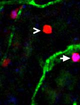
In our recent paper, Migrating pyramidal neurons require DSCAM to bypass the border of the developing cortical plate, we described the termination process of neuron radial migration and reported a molecular mechanism that underlies this process (Yang et al., 2022). We found that the nascent pyramidal neuron bypassing the last preceding neuron is important for expanding the cortex and thus determines the thickness of the upper cortical layers. Down syndrome cell adhesion molecule (DSCAM) was involved in this process by reducing the strength of N-cadherin-mediated cell-to-cell adhesion in the upper cortical plate, which allowed migrating neurons to traverse the cortical plate border and terminate at the outermost position of the developing cortical plate. Since DSCAM aberrations have been associated with autism spectrum disorder (Wang et al., 2016; Turner et al., 2016; Narita et al., 2020) and Down syndrome (Yamakawa et al., 1998; Agarwala et al., 2000), DSCAM may contribute to these brain disorders by impairing normal cortical development.
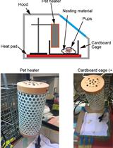
In our study, we applied live cell time-lapse imaging on organotypic brain-slice cultures to observe the termination of radial migration in detail (Yang et al., 2022). Organotypic brain slice cultures preserve brain development within an ex vivo environment. The confocal microscope is necessary for imaging fluorescently labeled neurons at single-cell resolution in thick brain slices. Since the brain slice is 300 µm in thickness, 50–100 z-frames of confocal scanning are needed to cover 100–150 µm of tissue in depth to collect z-axis information. Confocal microscopes equipped with high-speed scanners, such as the resonance scanner of Leica SP5 confocal systems, can overcome the speed limit of regular confocal microscopes and minimize laser damage. Together, this system generates single-cell resolution images for long-term (≥12 h) observations.
As we realized that our approach could be used as a standard method for studying cortical development and be extended to other brain regions or tissues, we decided to report our protocol in detail here, so that other researchers can reference, modify, and optimize their own imaging system. For the sake of completeness, we include brief protocols for neuronal labeling and brain slice sectioning in the “animal preparation” section, although these techniques have been previously described (Yang et al., 2022).
Materials and Reagents
15 mL Falcon tube for melting the agarose II (Corning, Falcon, catalog number: 14-959-53A)
3.5 × 1.0 cm Petri dish for mounting the fresh brain (Thermo Fisher Scientific, Nunc Petri Dish, catalog number: 150318)
Extra thick filter paper, precut (Bio-Rad, catalog number: 1703967)
10 cm Petri dish for carrying the new brain dissection (Thermo Fisher Scientific, Nunc Petri Dish, catalog number: 263991)
1/16’’, 3/16’’ inner diameter (i.d.) tubing to connect medium syringe to the sample chamber in the heater (ValveBank 4 II)
5/16’’ i.d. silicone tubing for bubbling
4 mm with medium-grit sandpaper (3M, catalog number: 9002NA-20-CC)
60 mL syringe
Transgenic mouse (Ai14, JAX, catalog number: 007914)
Agarose II (low melting point) (Amresco, catalog number: 17856)
2 L flask for carrying 1× artificial cerebral spinal fluid (ACSF)
Gas cylinder of 95% O2/5% CO2 gas
D-(+)-Glucose monohydrate (Millipore, catalog number: 49159)
NaCl (Fisher, catalog number: BP358-10)
KCl (Fisher, catalog number: BP366-1)
MgCl2·6H2O (Fisher, catalog number: BP214-500)
CaCl2·2H2O (Sigma, catalog number: 223506)
NaH2PO4·H2O (Sigma, catalog number: S9638)
NaHCO3 (Sigma, catalog number: S6014)
Deionized water (Millipore Milli-Q® Integral 5 Water Purification System, catalog number: ZRXQ005US)
Dulbecco’s modified Eagle medium (DMEM), high glucose (Sigma, catalog number: D0822)
Matrigel membrane matrix (Corning, Matrigel, catalog number: 356234)
10× ACSF solution I (see Recipes)
10× ACSF solution II (see Recipes)
1× ACSF (see Recipes)
Equipment
Electroporation (Harvard Apparatus, ECM 830)
Vibratome (Leica VT 1000)
ValveBank 4 II perfusion system (or similar systems)
Tissue culture incubator (37 °C, 5% CO2)
High pressure gas cylinder of 95% O2/5% CO2
Homemade sample dish modified from a 3.5 × 1.0 cm Petri dish. The height of the wall of the dish was reduced from 10 to 4 mm by grinding on a medium-grit sandpaper (e.g., 100 grit). The bottom of the dish was thinned by grinding on a fine-grit sandpaper (e.g., 600 grit).
Stage sample dish heater (Bioscience Tools, TC-E35)
Temperature controller (Bioscience Tools, TC-1-100)
Temperature fiber, miniature 0.87 mm (Bioscience Tools, TC-TP)
Silicon lid for the sample dish (Bioscience Tools, CSC-10P)
Vacuum oil pump (Fisher, VLP-200-115)
Confocal microscope (Leica SP5)
Software
ImageJ (NIH, https://imagej.nih.gov/ij/)
LAS X (Leica Microsystems, https://www.leica-microsystems.com/products/microscope-software/p/leica-las-x-ls/)
Procedure
文章信息
版权信息
© 2023 The Author(s); This is an open access article under the CC BY-NC license (https://creativecommons.org/licenses/by-nc/4.0/).
如何引用
Readers should cite both the Bio-protocol article and the original research article where this protocol was used:
- Yang, T., Hergenreder, T. and Ye, B. (2023). Analysis of Mouse Brain Sections by Live-cell Time-lapse Confocal Microscopy. Bio-protocol 13(7): e4648. DOI: 10.21769/BioProtoc.4648.
- Yang, T., Veling, M. W., Zhao, X. F., Prin, N. P., Zhu, L., Hergenreder, T., Liu, H., Liu, L., Rane, Z., Savelieff, M. G., et al. (2022). Migrating pyramidal neurons require DSCAM to bypass the border of the developing cortical plate. J Neurosci 42(28): 5510-5521.
分类
发育生物学 > 形态建成
神经科学 > 发育 > 形态建成
细胞生物学 > 细胞运动 > 细胞迁移
您对这篇实验方法有问题吗?
在此处发布您的问题,我们将邀请本文作者来回答。同时,我们会将您的问题发布到Bio-protocol Exchange,以便寻求社区成员的帮助。