

Multiphoton Microscopy of FITC-labelled Fusobacterium nucleatum in a Mouse in vivo Model of Breast Cancer
应用多光子显微镜检测小鼠体内乳腺癌模型中 FITC 标记的具核梭杆菌
发布: 2023年03月20日第13卷第6期 DOI: 10.21769/BioProtoc.4635 浏览次数: 1476
评审: Ivan ShapovalovAnonymous reviewer(s)
参见作者原研究论文
The authors used this protocol in:

Jun 2020
Advertisement


Abstract
Over the past decades, the main techniques used to visualize bacteria in tissue have improved but are still mainly based on indirect recognition of bacteria. Both microscopy and molecular recognition are being improved, but most procedures for bacteria detection in tissue involve extensive damage. Here, we describe a method to visualize bacteria in tissue slices from an in vivo model of breast cancer. This method allows examining trafficking and colonization of fluorescein-5-isothiocyanate (FITC)-stained bacteria in various tissues. The protocol provides direct visualization of fusobacterial colonization in breast cancer tissue. Rather than processing the tissue or confirming bacterial colonization by PCR or culture, the tissue is directly imaged using multiphoton microscopy. This direct visualization protocol causes no damage to the tissue; therefore, all structures can be identified. This method can be combined with others to co-visualize bacteria, types of cells, or protein expression in cells.
Background
Fusobacterium nucleatum is an oral anaerobic bacterium that is prevalent in periodontal disease (Nozawa et al., 2020) and preterm birth (Hill, 1993, 1998; Han et al., 2009; Gonzales-Marin et al., 2013). Recently, F. nucleatum was found to colonize colorectal adenocarcinoma (Castellarin et al., 2012; Kostic et al., 2012), esophageal cancer (Yamamura et al., 2016), pancreatic cancer (Mitsuhashi et al., 2015), and breast cancer (Parhi et al., 2020). The presence of F. nucleatum in colon, pancreatic, and esophageal cancer has been associated with poor prognosis (Mima et al., 2016; Yamamura et al., 2016; Yamaoka et al., 2018; Mitsuhashi et al., 2015). In a mouse model, F. nucleatum colonizes breast cancer and promotes tumor growth and metastatic progression (Parhi et al., 2020).
In order to identify Fusobacterium nucleatum colonization in human and mouse cancer tissues, the methods previously used were microbiome analysis, PCR, fluorescence in situ hybridization, and culture (Castellarin et al., 2012; Kostic et al., 2012; Parhi et al., 2020; Abed et al., 2016; Abed et al., 2020). Specifically directing bacteria to a target cancer as a future therapy strategy requires visualization tools for imaging and monitoring bacterial trafficking in animal models. Here, we show a simple method that requires minimal time, equipment, materials, and resources to quickly validate bacterial localization in cancer tissue in an in vivo model. This is a direct method to visualize the bacteria in the tissue with minimal intermediate steps that may affect the results.
Materials and Reagents
1 mL syringe (BD PlastipakTM, catalog number: 303084)
27G and 26G needle (BD PlastipakTM, catalog numbers: 305109 and 303176)
Sterile surgical pad
10 cm tissue culture dish (Corning Incorporated, Falcon, catalog number: 353003)
11 mL (NuncTM catalog number: 347856), 15 and 50 mL tubes (Miniplast, catalog numbers: 835-015-40-111 and 835-050-21-111)
1.5 mL Eppendorf microtubes (Bar Naor Ltd., catalog number: BN022RL)
Scalpel (Bar Naor Ltd., catalog number: BN400-21-JH)
Whatman syringe filter 0.2 µm (Bar Naor Ltd., catalog number: BNFCA206030H)
Coverslip (Bar Naor Ltd., catalog number: BN1001-18-1CN)
Superfrost Plus glass slides (Thermo Fisher Scientific, catalog number: J1800AMNT)
Pipette tips [Axygen, catalog numbers: TF-200-R-S (20–200 μL), TF-1000-R-S (100–1,000 μL)]
Glass vials (Bar Naor Ltd., catalog number: BN1228WH)
Animals: 5–7-weeks-old female C57BL/6 mice (Envigo, Israel)
AT3 cell line (Sigma SCC178)
Dulbecco’s modified eagle medium (DMEM), 500 mL (Biological Industries, catalog number: 01-050-1A)
Accutase solution normal human, 100 mL (Biological Industries, catalog number: C-41310)
Dulbecco’s PBS, 500 mL (Biological Industries, catalog number: 02-020-1A)
Heat-inactivated fetal bovine serum (FBS), 500 mL (Biological Industries, catalog number: 04-127-1A)
L-glutamine in saline solution, 100 mL (Biological Industries, catalog number: 03-020-1B)
Penicillin–streptomycin solution, 100 mL (Biological Industries, catalog number: 03-031-1B)
MEM non-essential amino acids solution, 100 mL (Biological Industries, catalog number: 01-340-1B)
Fusobacterium nucleatum strain of interest (for example ATCC 23726)
Wilkins-Chalgren broth (Oxoid, catalog number: CM0643)
Fluorescein isothiocyanate (FITC), 1 g (Sigma-Aldrich, catalog number: F7250-1G)
Ethanol (Sigma-Aldrich, catalog number: E7023)
Trypan blue solution (Biological Industries, catalog number: 03-102-1B)
Sodium carbonate (Sigma-Aldrich, catalog number: 497-19-8)
Sodium hydroxide (Sigma-Aldrich, catalog number: 1310-73-2)
Isoflurane, USP TerrellTM (Piramal, catalog number: VED1350)
Double deionized water (DDW)
Ice
Wilkins medium (see Recipes)
Sodium carbonate buffer (0.1 M, pH 9.0) (see Recipes)
FITC solution (see Recipes)
Equipment
Pipettes
Centrifuge (Eppendorf 5810 R)
Autoclave (Tuttnauer 2540MK)
Anesthesia induction chamber
Hemocytometer
Spectrophotometer (600 nm wavelength)
Orbital shaker
Nikon multiphoton A1MP
Stereo microscope (Nikon SMZ2-)
Scalpel handle
Scissors
Forceps
Tail vein injection platform
Heat lamp
Anaerobic chamber (Bactron I–II Shellab, USA)
Caliper
Vortex
Software
NIS-Elements (Nikon Instruments Inc.) https://www.microscope.healthcare.nikon.com/products/software/nis-elements)
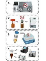
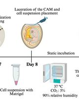
Procedure
文章信息
版权信息
© 2023 The Authors; exclusive licensee Bio-protocol LLC.
如何引用
Parhi, L., Shhadeh, A., Maalouf, N., Alon-Maimon, T., Scaiewicz, V. and Bachrach, G. (2023). Multiphoton Microscopy of FITC-labelled Fusobacterium nucleatum in a Mouse in vivo Model of Breast Cancer. Bio-protocol 13(6): e4635. DOI: 10.21769/BioProtoc.4635.
分类
癌症生物学 > 通用技术 > 动物模型
生物科学 > 微生物学 > 微生物群落
您对这篇实验方法有问题吗?
在此处发布您的问题,我们将邀请本文作者来回答。同时,我们会将您的问题发布到Bio-protocol Exchange,以便寻求社区成员的帮助。