

Visualization of Lipid Droplets in the Alveolar Macrophage Cell Line MH-S with Live-cell Imaging by 3D Holotomographic Microscopy (Nanolive)
通过 3D 全息显微术 (Nanolive)的活细胞成像实现肺泡巨噬细胞系 MH-S 中脂滴的可视化


发布: 2023年03月05日第13卷第5期 DOI: 10.21769/BioProtoc.4629 浏览次数: 1807
评审: David PaulVasiliki KoliarakiAnonymous reviewer(s)
参见作者原研究论文
The authors used this protocol in:

Feb 2022
Advertisement


Abstract
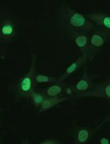
Lipid droplets (LD), triglycerides and sterol esters among them, are well known for their capacity as lipid storage organelles. Recently, they have emerged as critical cytoplasmic structures involved in numerous biological functions. LD storage is generated de novo by the cell and provides an energy reserve, lipid precursors, and cell protection. Moreover, LD accumulation can be observed in some pathologies as obesity, atherosclerosis, or lung diseases. Fluorescence imaging techniques are the most widely used techniques to visualize cellular compartments in live cells, including LD. Nevertheless, presence of fluorophores can damage subcellular components and induce cytotoxicity, or even alter the dynamics of the organelles. As an alternative to fluorescence microscopy, label-free techniques such as stimulated Raman scattering and coherent anti-stokes Raman scattering microscopy offer a solution to avoid the undesirable effects caused by dyes and fluorescent proteins, but are expensive and complex. Here, we describe a label-free method using live-cell imaging by 3D holotomographic microscopy (Nanolive) to visualize LD accumulation in the MH-S alveolar macrophage cell line after treatment with oleic acid, a monounsaturated fatty acid that promotes lipid accumulation.
Keywords: Lipid droplet (脂滴)Background
Lipid droplets (LD) are dynamic cytoplasmic organelles that serve as intracellular energy storage, mainly in the form of triglycerides and cholesterol esters (van Dierendonck et al., 2022). In addition to their role in lipid storage and transport, LD are increasingly recognized to be involved in the regulation of other cellular processes, including inflammation, cell activation, and metabolism (Xu et al., 2018; Agudelo et al., 2020). Accumulation of LD is also related to several pathologies as obesity, diabetes, atherosclerosis, or lung diseases (Xu et al., 2018; Agudelo et al., 2020). Alveolar macrophages (AM) are the first line of defense against respiratory pathogens and play key roles in lung lipid metabolism (Agudelo et al., 2020). Excessive amounts of intracellular lipids in AM have been described in lung pathologies such as chronic obstructive pulmonary disease, acute lung injury, idiopathic pulmonary fibrosis, or pulmonary alveolar proteinosis, among others (Agudelo et al., 2020). Nevertheless, the mechanisms responsible for LD accumulation are not fully elucidated. Fluorescence-based live-cell imaging is the most widely used technique for LD studies, even though the phototoxicity of fluorescent dyes may disturb LD dynamics. Here, we present a protocol for live imaging of the LD accumulation without the addition of fluorescent labels, using 3D holotomographic microscopy (Nanolive). This label-free microscopy method reports the changes of the refractive indices (RIs) in three dimensions at high spatial and temporal resolution, allowing label-free analysis of organelle biology and kinetics studies with a low level of phototoxicity (Sandoz et al., 2019). We have performed this protocol in the MH-S alveolar macrophage cell line, grown in the presence of oleic acid to induce LD formation. Using this live-cell approach, foam cell formation can be analyzed under a physiological context, excluding the artefacts induced by the addition of different chemicals.
Materials and Reagents
µ-dish cell culture imaging dish, 35 mm, high (Ibidi, catalog number: 81156)
1,000, 200, and 20 µL pipette tips (pre-sterile w/ filter, hinged racks) (ExpellPlus, catalog numbers: 5030150C, 5030090C, 5130062C)
15 mL centrifuge tubes (Corning, catalog numbers: 430791)
Corning® Costar® Stripette® serological pipettes, individually paper/plastic wrapped, 5 and 10 mL (Corning, catalog numbers: CLS4487, 4488)
Falcon® 100 mm TC-treated cell culture dish (Corning, catalog number: 353003)
RPMI 1640 media (Lonza, catalog number: BE 12-115F)
Fetal bovine serum (FBS) (GibcoTM, catalog number: 10270106)
Penicillin-streptomycin mixture (Lonza, catalog number: DE17-603E)
Dulbecco's phosphate buffered saline (PBS) (10×), 95 mM (PO4) without calcium or magnesium (Lonza, catalog number: BE17-515Q)
Bovine serum albumin (BSA)-oleate monounsaturated fatty acid complex (5 mM) (Cayman Chemical, catalog number: 29557)
BSA control for BSA-fatty acid complexes (5 mM) (Cayman Chemical, catalog number: 29556)
Trypsin/EDTA solution (Lonza, catalog number: CC-5012)
Complete RPMI growth medium (see Recipes)
Starving medium (see Recipes)
Biological materials
Cell line: MH-S (ATCC number: CRL-2019TM)
Equipment
Rainin Pipet-Lite XLS (Mettler Toledo, models: SL1000 and SL200)
Forma Direct heat CO2 incubator 184 L digital model (Thermo Scientific, model: 311; TC 230)
Biosafety cabinet (Telstar, model: Bio II Advance Plus IV)
Centrifuge Sorvall ST 16R (Thermo Scientific, model: 75004380)
3D Cell Explorer microscope (Nanolive, Ecublens, Switzerland)
CellDropTM FL automated cell counter (DeNovix)
Laboratory water bath (Memmert, model: WNB 14)
Software
Steve software v1.6.3496 (Nanolive, Ecublens, Switzerland)
ImageJ (FIJI) software (https://imagej.net/software/fiji/downloads)
Procedure
文章信息
版权信息
© 2023 The Author(s); This is an open access article under the CC BY-NC license (https://creativecommons.org/licenses/by-nc/4.0/).
如何引用
Pérez-Montero, A., Zaragoza, O., Luque, A., Hortelano, S. and Acebo, P. (2023). Visualization of Lipid Droplets in the Alveolar Macrophage Cell Line MH-S with Live-cell Imaging by 3D Holotomographic Microscopy (Nanolive). Bio-protocol 13(5): e4629. DOI: 10.21769/BioProtoc.4629.
分类
生物物理学 > 显微技术
细胞生物学 > 细胞成像 > 活细胞成像
您对这篇实验方法有问题吗?
在此处发布您的问题,我们将邀请本文作者来回答。同时,我们会将您的问题发布到Bio-protocol Exchange,以便寻求社区成员的帮助。