

Laser Capture Microdissection (LCM) of Human Skin Sample for Spatial Proteomics Research
用于空间蛋白质组学研究的人体皮肤样本激光捕获显微切割(LCM)
(*contributed equally to this work) 发布: 2023年03月05日第13卷第5期 DOI: 10.21769/BioProtoc.4623 浏览次数: 2271
评审: Zinan ZhouDarrell CockburnAnonymous reviewer(s)
参见作者原研究论文
The authors used this protocol in:

Jul 2022
Advertisement


Abstract
In mammals, the skin comprises several distinct cell populations that are organized into the following layers: epidermis (stratum corneum, stratum granulosum, stratum spinosum, and basal layer), basement membrane, dermis, and hypodermal (subcutaneous fat) layers. It is vital to identify the exact location and function of proteins in different skin layers. Laser capture microdissection (LCM) is an effective technique for obtaining pure cell populations from complex tissue sections for disease-specific genomic and proteomic analysis. In this study, we used LCM to isolate different skin layers, constructed a stratified developmental lineage proteome map of human skin that incorporates spatial protein distribution, and obtained new insights into the role of extracellular matrix (ECM) on stem cell regulation.
Keywords: Skin (皮肤)Background
Skin, the largest barrier organ in the body, has a tough and pliable structure to adapt to external conditions by quickly repairing mechanical, chemical, and biological injuries. In mammals, the skin consists of several distinct layers: the epidermal, dermal, and subcutaneous fat layers. The epidermis is the outermost layer, with stratified cell layers maintained by keratinocytes including stem cells and an abundance of mature cells (Gonzales and Fuchs, 2017). The basal layers of the epidermis, which express the keratins KRT5 and KRT14, are the location of undifferentiated proliferative epidermal stem cells (Blanpain and Fuchs, 2006). The progenitor cells replenish basal layers and differentiate into mature keratinocytes in the granulosum and spinosum layers, which express KRT1, KRT10, and involucrin. Stratum corneum, the outermost layer, consisting of terminally differentiated and dead cells, acts as a scaffold for the lipid bilayers that comprise the epidermal barrier on the skin surface (Fuchs, 2007; Koster and Roop, 2007). Keratins, which constitute the cytoskeleton of epithelial cells, are highly expressed in the epidermis, especially in the stratum corneum (Li et al., 2022). In the epidermis, the transition of keratinocytes from the proliferative basal to the supra-basal cell layer during terminal differentiation and keratinization is characterized by keratin expression transition from basal cell keratins (KRT5, KRT14, and KRT15) to supra-basal cell keratins (KRT1 and KRT10) (Moll et al., 2008).The dermis provides nourishment and support for the skin and contains an abundance of extracellular matrix (ECM) proteins including collagens, glycosaminoglycans, hyaluronic acid, fibronectin, elastin, and laminin (Nyström and Bruckner-Tuderman, 2019). Basement membrane, a specialized layer of ECM proteins connecting the dermis and epidermis, serves as an important microenvironment for basal stem cells; understanding its composition is crucial to the study of basal stem cell fate and function. However, there is a lack of detailed information about the molecular composition and regulatory function of specialized proteins localized in different skin layers that are difficult to separate, especially the basement membrane zone, during the dynamic processes of skin development, homeostasis, and regeneration for wound healing (Dengjel et al., 2020; Dyring-Andersen et al., 2020).
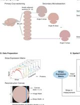
As described above, the skin is a complex tissue composed of heterogeneous cell types with different spatial distributions. Studying the unique physiological and pathological functions of different spatially distributed cell types is essential for understanding the molecular characteristics of human skin. Laser capture microdissection (LCM) is a powerful technique for separating target cell populations with extremely high microscopic precision, thus providing a perfect solution to the problem of skin tissue heterogeneity. Here, we describe a protocol for the isolation of different skin layers by LCM for spatial proteomics research (Li et al., 2022). Using a combination of previously developed tissue engineering decellularization methods, dedicated to the removal of epidermis and separation of basement membrane (Leng et al., 2020, Liu et al., 2020), LCM, and mass spectrometry (MS), we isolated proteins from six skin layers (stratum corneum, stratum granular-spinous, basal layer, basement membrane, superficial dermis, and deep dermis) and constructed a stratified developmental lineage proteome map of human skin. By obtaining different skin samples, the protocol enables the analysis of spatial protein expression in normal and disease-specific skin tissues, which is important for understanding the pathogenesis of different skin diseases and discovering potential therapeutic targets.
Materials and Reagents
MMI MembraneSlidesTM (MMI GmbH, catalog number: 50102)
MMI IsolationCaps transparent, 0.5 mL (MMI GmbH, catalog number: 50204)
Phospholipase A2 (Sigma-Aldrich, catalog number: P6534)
Sodium deoxycholate (Sigma-Aldrich, catalog number: V900388)
PBS (TBD Science, catalog number: PB2004Y)
EDTA (Sigma-Aldrich, catalog number: E9884)
NaCl (Sigma-Aldrich, catalog number: S9888)
DNase (Sigma-Aldrich, catalog number: 11284932001)
RNase (Sigma-Aldrich, catalog number: 10109134001)
OCT compound (SAKURA, catalog number: 4583)
4% paraformaldehyde (BOSTER Biological Technology, catalog number: AR1068)
Sucrose (Sigma-Aldrich, catalog number: V900116)
Delipidation solution (see Recipes)
DNase-RNase solution (see Recipes)
3.4 M NaCl solution (see Recipes)
30% sucrose solution (see Recipes)
Equipment
Laser microdissection system (MMI, cellcut plus)
Freezing microtome (Leica, model: CM1950)
Shaker (tcsysb, THZ-C)
Scalpel (BELEVOR MEDICAL, catalog number: 03.0030.01)
Procedure
文章信息
版权信息
© 2023 The Author(s); This is an open access article under the CC BY-NC license (https://creativecommons.org/licenses/by-nc/4.0/).
如何引用
Zhang, Q., Gong, H., Ma, J., Li, J. and Leng, L. (2023). Laser Capture Microdissection (LCM) of Human Skin Sample for Spatial Proteomics Research. Bio-protocol 13(5): e4623. DOI: 10.21769/BioProtoc.4623.
分类
细胞生物学 > 组织分析 > 组织分离
生物化学 > 蛋白质 > 定量
干细胞 > 成体干细胞 > 上皮干细胞
您对这篇实验方法有问题吗?
在此处发布您的问题,我们将邀请本文作者来回答。同时,我们会将您的问题发布到Bio-protocol Exchange,以便寻求社区成员的帮助。