

Comprehensive Analyses of Muscle Function, Lean and Muscle Mass, and Myofiber Typing in Mice
小鼠肌肉功能、瘦肌肉质量以及肌纤维分型的综合分析
(*contributed equally to this work) 发布: 2023年02月20日第13卷第4期 DOI: 10.21769/BioProtoc.4617 浏览次数: 3300
评审: Vivien Jane Coulson-ThomasAllan-Hermann PoolNeha NandwaniAbhilash PadavannilAnonymous reviewer(s)
参见作者原研究论文
The authors used this protocol in:

Feb 2022


Abstract
Skeletal muscle disorders commonly affect the function and integrity of muscles. Novel interventions bring new potential to rescue or alleviate the symptoms associated with these disorders. In vivo and in vitro testing in mouse models allows quantitative evaluation of the degree of muscle dysfunction, and therefore, the level of potential rescue/restoration by the target intervention. Several resources and methods are available to assess muscle function and lean and muscle mass, as well as myofiber typing as separate concepts; however, a technical resource unifying these methods is missing. Here, we provide detailed procedures for analyzing muscle function, lean and muscle mass, and myofiber typing in a comprehensive technical resource paper.
Graphical abstract

Background
Skeletal muscle homeostasis and dysfunction depend on the adaptative changes in myofiber size, myofiber type, and force capacity. Equally important for both disease characterization and therapeutic evaluation are sensitive and precise methods for evaluating the parameters of muscle physiology in comprehensive assessments. This set of tools will enable scientists to assess not only the magnitude of the effect of a target intervention but also compare effects across different interventions.
Multiple invasive and noninvasive methods have been developed to assess muscle strength in animal models, especially rodents (Takeshita et al., 2017). Invasive methods include in situ and in vitro measurement of muscle force, whereas noninvasive methods include in vivo assessment of strength and global performance. In this protocol, we present the methods for conducting both in situ invasive measurements of muscle force and in vivo non-invasive measurements of strength and aerobic exercise tolerance in response to a pro-ergogenic glucocorticoid treatment (Quattrocelli et al., 2022).
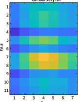
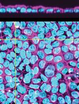
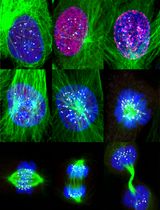
Skeletal muscle is a metabolically active tissue made up of different types of muscle fibers, generally classified as type1, type 2A, type 2X, and type 2B according to the myosin isoform that is enriched in each of them (Talbot and Maves, 2016). Changes in muscle fiber typing are often correlated with changes in muscle strength, contractility, and/or endurance. Moreover, modifications in skeletal muscle fiber composition may be related to muscle myopathies such as muscular dystrophies, inherited myopathies preferentially affecting a specific myofiber type, and sarcopenia. Therefore, myofiber-type specification is important to provide insights for understanding the susceptibility and resistance of certain fiber types to muscle disease and to discover potential treatments based on muscle plasticity and metabolic shift. Histochemical staining for myosin ATPase or succinate dehydrogenase enzyme activity or quantification of metabolic enzymes are among the methods used to identify different myofiber types, but these cannot generally differentiate between myofibers subtypes (Reichmann and Pette, 1984). Therefore, here we report the immunofluorescence-based protocol that is routinely used in our lab for the differential staining of myosin heavy chain isoforms, which efficiently resolves the heterogeneity of muscle fibers.
Apart from muscle function and myofiber typing, lean mass (Kalyani et al., 2014) and muscle mass (Lovering et al., 2005) are two important determinants associated with muscle disorders or muscle changes downstream of global conditions, such as aging or type-2 diabetes. Weight curves offer an immediate proxy estimate of overall growth and mass levels but evidently cannot provide information on fat vs. lean mass or actual muscle mass. Therefore, magnetic resonance imaging (MRI)-based systems to estimate lean and fat mass can provide a better estimation of body composition effects of interventions, such as circadian time–specific dosing of glucocorticoids (Quattrocelli et al., 2022). Using MRI-based systems, it is possible to simultaneously quantify changes in adiposity vs. lean mass (absolute or relative) over time.
Overall, we provide a comprehensive method for the analysis of muscle function, performance, lean and muscle mass, and muscle fiber typing. These tests can be easily customized to evaluate overall muscle function, performance, and composition (especially in terms of muscle fiber types) in different situations, to study muscle physiopathology, or to assess the efficacy of therapeutic interventions.
Materials and Reagents
Microscope slides (Corning, catalog number: CLS294775X25)
Microscope slide cover glass (Globe Scientific, catalog number: 1404-10)
Phosphate buffered saline (PBS) (Sigma-Aldrich, catalog number: 806552)
Triton X-100 (Sigma-Aldrich, catalog number: T8787)
10% normal goat serum (Cell Signaling, catalog number: 5425)
Ethanol, 200 proof (Thermo Scientific, catalog number: T038181000)
Isoflurane (Covetrus, catalog number: 11695-6777-2)
Ringer’s solution (Fisher Scientific, catalog number: S25512)
Isopentane (Alfa Aesar, catalog number: 19387, CAS number: 78-78-4)
Tissue-Tek OCT compound (Sakura Finetek, catalog number: 4583)
PAP pen (Biotium, catalog number: 22006)
ProLongTM Gold Antifade Mountant with DAPI (Invitrogen, catalog number: P36931)
Primary antibodies (Table 1)
Table 1. Primary antibodies for muscle fiber typing
Antibody name Company and catalog number Dilution Rabbit anti(α)-Laminin Sigma, L9393 1:500 Myosin heavy chain, type IIA (SC-71) Developmental Studies Hybridoma Bank, SC-71 1:30 Myosin heavy chain, type IIB (BF-F3) Developmental Studies Hybridoma Bank, 10F5 1:10 Myosin heavy chain (slow) (BA-F8) Developmental Studies Hybridoma Bank, A4.840 1:10 Fluorescently tagged secondary antibodies (Table 2)
Table 2. Secondary antibodies for muscle fiber typing
Antibody Name Company and Cat No. Dilution Goat anti-rabbit IgG (H+L) Alexa FluorTM 488 Invitrogen, A-11034 1:1,000 Goat anti-mouse IgG2b Alexa FluorTM 647 Invitrogen, A-21242 1:1,000 Goat anti-mouse IgG1, Alexa FluorTM 488 Invitrogen, A21121 1:1,000 Goat anti-mouse IgM, Alexa FluorTM 594 Invitrogen, A-21044 1:1,000 Goat serum (Invitrogen, catalog number: 31872)
Dimethyl sulfoxide (DMSO) (Fisher Scientific, catalog number: D1319)
Prednisone powder (Sigma-Aldrich, catalog number: P6254)
1 mL insulin syringe with needle (BD, catalog number: 329424)
Permeabilization reagent: 1% (v/v) Triton X-100 in PBS
Blocking buffer: 10% (v/v) goat serum in PBS
Prednisone stock (5 mg/mL): 5 mg prednisone powder in 1 mL DMSO
Physiological solution: sterile 0.9% saline solution (Cytiva Z1379)
Vehicle stock: 1 mL DMSO.
Equipment
Grip and treadmill assays
Grip strength meter with single sensor for mice (max 1 kg capacity) with standard pull bars (Chatillon Instruments, Digital Force Gauge, catalog number: DFIS-2)
Open treadmill with stimulus assembly, ~10% uphill incline, individual mouse lanes, drive motor to adjust speed (3–100 m/min range), and exercising belt with grip-prone texture (Columbus Instruments, catalog number: Exer3/6)
Force-frequency analysis in tibialis anterior muscles
Surgical scissors (Fine Science Tools, catalog number: 14060-10)
Mouse handling forceps (Fine Science Tools, catalog number: 11036-20)
Weighing scale (Ohaus, catalog number: 80000022)
In-situ apparatus setup from 3-in-1 whole animal system for mice (Aurora Scientific, catalog number: 1300A)
Ohaus Pioneer precision balance (Fisher Scientific, catalog number: 01-922-178)
Digital caliper (Thermo Fisher, catalog number: 14-648-17)
Isoflurane vaporizer (SurgiVet Classic T3, catalog number VCT302) and induction chamber (Patterson Scientific, catalog number: 07-8917760)
Surgical suture (Ethicon, catalog number: K871)
Myofiber typing
Leica cryostat microtome (Leica Biosystems, catalog number: CM1510S)
Humidified container (Thomas Scientific, catalog number: 1219D68)
Nikon Eclipse Ti inverted microscope (Nikon Instruments, catalog number: Ti-E)
MRI scans to determine lean mass ratios
EchoMRI whole-body composition analyzer (EchoMRI, catalog number: 100H)
Sample tubes dedicated to mice comprised between 20 and 40 g of body mass (EchoMRI, Houston, TX)
Standard internal calibrator tube (canola oil) (EchoMRI, Houston, TX)
Software
Built-in software EchoMRI version 140320. Data were analyzed when hydration ratio was >85%
ImageJ (Fiji, https://imagej.nih.gov/ji)
ASI 611A Dynamic Muscle Analysis v5.300
ASI 610A Dynamic Muscle Control v5.500
Procedure
文章信息
版权信息
© 2023 The Author(s); This is an open access article under the CC BY-NC license (https://creativecommons.org/licenses/by-nc/4.0/).
如何引用
Readers should cite both the Bio-protocol article and the original research article where this protocol was used:
- Durumutla, H. B., Villa, C., Panta, M., Wintzinger, M., Pragasam, A. D., Miz, K. and Quattrocelli, M. (2023). Comprehensive Analyses of Muscle Function, Lean and Muscle Mass, and Myofiber Typing in Mice. Bio-protocol 13(4): e4617. DOI: 10.21769/BioProtoc.4617.
- Quattrocelli, M., Wintzinger, M., Miz, K., Levine, D. C., Peek, C. B., Bass, J. and McNally, E. M. (2022). Muscle mitochondrial remodeling by intermittent glucocorticoid drugs requires an intact circadian clock and muscle PGC1alpha. Sci Adv 8(7): eabm1189.
分类
生物科学 > 生物技术
细胞生物学 > 细胞成像 > 固定细胞成像
您对这篇实验方法有问题吗?
在此处发布您的问题,我们将邀请本文作者来回答。同时,我们会将您的问题发布到Bio-protocol Exchange,以便寻求社区成员的帮助。
![]() 提问指南
提问指南
+ 问题描述
写下详细的问题描述,包括所有有助于他人回答您问题的信息(例如实验过程、条件和相关图像等)。