

Lysate-to-grid: Rapid Isolation of Native Complexes from Budding Yeast for Cryo-EM Imaging
裂解液到网格:从出芽酵母中快速分离天然复合物用于低温冷冻电镜成像
发布: 2023年01月20日第13卷第2期 DOI: 10.21769/BioProtoc.4596 浏览次数: 2915
评审: Laxmi Narayan MishraSashikantha Reddy PulikalluNityanand Srivastava
参见作者原研究论文
The authors used this protocol in:
Aug 2019


Abstract
Single-particle electron cryo-microscopy (cryo-EM) is an effective tool to determine high-resolution structures of macromolecular complexes. Its lower requirements for sample concentration and purity make it an accessible method to determine structures of low-abundant protein complexes, such as those isolated from native sources. While there are many approaches to protein purification for cryo-EM, attaining suitable particle quality and abundance is generally the major bottleneck to the typical single-particle project workflow. Here, we present a protocol using budding yeast (S. cerevisiae), in which a tractable immunoprecipitation tag (3xFLAG) is appended at the endogenous locus of a gene of interest (GOI). The modified gene is expressed under its endogenous promoter, and cells are grown and harvested using standard procedures. Our protocol describes the steps in which the tagged proteins and their associated complexes are isolated within three hours of thawing cell lysates, after which the recovered proteins are used directly for cryo-EM specimen preparation. The prioritization of speed maximizes the ability to recover intact, scarce complexes. The protocol is generalizable to soluble yeast proteins that tolerate C-terminal epitope tags.
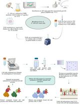
Graphical abstract

Overview of lysate-to-grid workflow. Yeast cells are transformed to express a tractable tag on a gene of interest. Following cell culture and lysis, particles of interest are rapidly isolated by co-immunoprecipitation and prepared for cryo-EM imaging (created with BioRender.com).
Background
Structural determination of biological macromolecules is essential for providing insights into their function. Over the past decade, revolutionary advances in hardware and software have ushered in a new era of single-particle electron cryo-microscopy (cryo-EM) (Kühlbrandt, 2014; Shen, 2017). The field is amidst a golden age, as evidenced by recent structures that have achieved atomic resolution (Nakane et al., 2020; Yip et al., 2020). Such resolution has traditionally only been accessible through other established methods, such as X-ray crystallography and nuclear magnetic resonance, which also generally require a substantial amount of material (e.g., on the order of several milligrams). In contrast, single particle cryo-EM only requires enough material to cover the support grid, and a few microliters of sample between 0.1 and 1.0 micromolar concentration is often sufficient for each specimen.
Common approaches to generate sufficient material for structural studies typically depend on overexpressing proteins in heterologous systems. While overexpression systems are effective in producing large quantities of material, the purified proteins may lack relevant binding partners or post-translational modifications that do not exist in such systems. The relatively low sample volume and concentration requirements for cryo-EM make it an attractive method to determine structures of protein complexes that are isolated directly from their native sources. Indeed, cryo-EM imaging of proteins from cell extracts is increasingly becoming more feasible as a tool for discovery biology (Ho et al., 2020; Skalidis et al., 2022). Such top-down approaches enable the characterization of particles in a more native-like context that is inherently missing in bottom-up in vitro reconstitution methods. Yet, achieving sufficient material of scarce or short-lived complexes remains a major barrier to preparing high-quality cryo-EM specimens.
By leveraging the advantages of working with the budding yeast model organism (S. cerevisiae), including genetic tractability and throughput of cell growth, we have optimized a generalizable procedure that enables the rapid isolation of endogenous complexes from cell lysates. The procedure balances between speed and purity, and samples are ready for cryo-EM specimen vitrification within three hours. The prioritization of speed enables the recovery of multi-component complexes that may otherwise fall apart in lengthier procedures, such as those with overnight steps. Our procedure was used to isolate scarce complexes trapped in their active, functional states, including capturing the processes of mRNA-independent peptide synthesis by the ribosome-associated quality control complex (~300 copies per cell; Shen et al., 2015) and protein unfolding by the Shp1-Cdc48 AAA+ ATPase complex (~3,000 copies per cell; Cooney et al., 2019; protein copy numbers estimated from Ghaemmaghami et al., 2003). In both of these studies, the rapid isolation of native complexes enabled the structure determination of multi-component complexes in novel and biologically important functional states.
Here, we describe our detailed protocol of how native multi-component, soluble complexes are quickly isolated from yeast lysates and used for cryo-EM imaging. A C-terminal 3xFLAG tag is inserted into the endogenous locus of a gene of interest (GOI) via homologous recombination. Cells are grown and harvested and their lysates are prepared for co-immunoprecipitation (co-IP) experiments. The target protein and its associated complexes are recovered by competitive elution and used directly for cryo-EM specimen preparation. In principle, our protocol can be applied to isolate and image any soluble yeast protein and their associated complexes as long as their function is not disrupted by the C-terminal tag and the particles are of sufficient size to be visible by cryo-EM.
Materials and Reagents
General supplies
250 mL Erlenmeyer flasks (Corning, catalog number: 4980-250)
2.8 L Fernbach culture Pyrex flasks (Sigma-Aldrich, catalog number: CLS44202XL)
1.5 mL Eppendorf/microcentrifuge tubes (Fisher Scientific, catalog number: 14-282-302)
15 mL conical tubes (Greiner Bio-One, catalog number: 188271)
50 mL conical tubes (Greiner Bio-One, catalog number: 227270)
15 mL glass vials (Corning, catalog number: 9820-16X)
Small growth platform shaker (Lab-Line Instruments, Inc., catalog number: 3590-1)
Large growth platform shaker (Barnstead | Lab-Line, A-Class., catalog number: SHKA3000)
1 L centrifuge bottle and caps (Beckman Coulter, catalog number: C31597)
Styrofoam box
Membrane filter, 0.22 μm pore size (Millipore, catalog number: GSWP04700)
pFA6a plasmid (Longtine et al., 1998). We use pTF272 (pFA6a-TEV-6xGly-3xFLAG-HphMX) (Addgene plasmid # 44083; http://n2t.net/addgene:44083; RRID: Addgene_44083)
iProof HF 2X Master mix (Bio-Rad, catalog number: 1725310)
GeneRuler 1 kb Plus DNA ladder (ThermoFisher Scientific, catalog number: SM1331)
EZVision One Dye-as-Loading buffer, 6× (VWR, catalog number: 97064-190)
Agarose (GoldBio, catalog number: A-201-500)
Ethylenediaminetetraacetic acid (EDTA) (Fisher Scientific, catalog number: AC118432500)
Acetic acid (Fisher Chemical, catalog number: BP2401C-212)
S. cerevisiae yeast strain BY4741 (derived from S288C strain; genotype MATa; his3Δ1; leu2Δ0; met15Δ0; ura3Δ0)
Yeast extract (Fisher Scientific, catalog number: BP9727-2)
Peptone (Fisher Scientific, catalog number: BP9725)
Adenine (Sigma-Aldrich, catalog number: A8626)
Glucose (Sigma-Aldrich, catalog number: G7021)
Agar (Becton Dickson & Company, catalog number: 2013-03-31)
Poly(ethylene glycol) (PEG) 3350 (Sigma-Aldrich, catalog number: 202444)
Sheared salmon sperm DNA (Ambion, catalog number: 1009027)
Lithium acetate (Sigma-Aldrich, catalog number: 517992)
Hygromycin B (Sigma-Aldrich, catalog number: 10843555001)
Sodium dodecyl sulfate (SDS) (Fisher Scientific, catalog number: BP166)
4%–15% precast protein gel (Bio-Rad, catalog number: 4561086)
2× Laemmli sample buffer (Bio-Rad, catalog number: 1610737)
Beta-mercaptoethanol (Bio-Rad, catalog number: 161-0710)
Methanol (Fisher Scientific, catalog number: A412P-4)
PVDF membrane (Bio-Rad, catalog number: 1620177)
Precision plus protein dual color standards (Bio-Rad, catalog number: 161-0374)
Thick blot filter paper (Bio-Rad, catalog number: 1703969)
Roller (ThermoFisher Scientific, catalog number: 0084747)
Skim milk powder (MP Bio, catalog number: 902887)
Tween 20 (Sigma-Aldrich, catalog number: 11332465001)
Incubation box (Li-Cor, catalog number: 929-97301)
Monoclonal ANTI-FLAG® M2 antibody produced in mouse (Sigma-Aldrich, catalog number: F1804)
Goat anti-mouse IgG secondary antibody (Li-Cor, catalog number: 926-32210)
Sodium azide (Sigma-Aldrich, Catalog number: S2002)
Glycerol (Fisher Bioreagents, catalog number: BP229)
HEPES (Fisher Bioreagents, catalog number: BP310-500)
Potassium acetate (KOAc) (Sigma-Aldrich, catalog number: P1190)
Magnesium acetate [Mg(Oac)2] (Sigma-Aldrich, catalog number:M5661)
Calcium chloride (CaCl2) (Fisher Chemical, catalog number: C79-500)
D-sorbitol (Sigma-Aldrich, catalog number: 1003101761)
Phenylmethylsulfonyl fluoride (PMSF) (Sigma-Aldrich, catalog number: P7626)
Aprotinin (GoldBio, catalog number: A-655)
Pepstatin (GoldBio, catalog number: P-020)
Leupeptin (GoldBio, catalog number L-010)
Dithiothreitol (DTT) (GoldBio, catalog number DTT)
Liquid nitrogen
Magnesium chloride hexahydrate (MgCl2·6H2O) (Acros Organics, catalog number: 41341-500)
Igepal CA-630 (Sigma-Aldrich, catalog number I3021)
Anti-FLAG M2 affinity gel (Sigma-Aldrich, catalog number: A2220)
3xFLAG peptide (APExBio, catalog number: A 6001)
Formvar/Carbon 200 mesh grids (Ted Pella, Inc., catalog number: 01801)
595 Vitrobot filter paper (Ted Pella, Inc., catalog number: 47000-100)
Uranyl acetate salt (EMS, catalog number: 22400)
Glutaraldehyde (EMS, Catalog number: 16000)
Tris hydrochloride (Tris-HCl) (Sigma-Aldrich, Catalog number: 9310-OP)
Quantifoil R1.2/1.3 or UltrAuFoil R1.2/1.3 300 mesh grids (SPT Labtech)
Compressed nitrogen gas cylinder
Compressed ethane gas cylinder
Stock solutions
50× TAE (see Recipes)
5× TBS (see Recipes)
10× transfer buffer (see Recipes)
1 M HEPES-KOH, pH 7.4 (see Recipes)
1 M KOAc (see Recipes)
1 M Mg(Oac)2 (see Recipes)
1 M CaCl2 (see Recipes)
1 M sorbitol (see Recipes)
1 M MgCl2 (see Recipes)
1 M DTT (see Recipes)
2× IP buffer (see Recipes)
1 M lithium acetate (LiOAc) (see Recipes)
Gels
1% agarose gel (see Recipes)
Buffers
1× TAE (see Recipes)
Working transfer buffer (see Recipes)
Blocking solution (see Recipes)
TBST solution (see Recipes)
Yeast lysis buffer (see Recipes)
Wash buffer 1 (see Recipes)
Wash buffer 2 (see Recipes)
Mixes
Transformation PCR mix (according to Bio-Rad iProof HF manual) (see Recipes)
Colony PCR mix (see Recipes)
PEG mix (see Recipes)
Reagents
Lithium acetate (LiOAc) (see Recipes)
1% uranyl acetate (see Recipes)
Tris base (Sigma-Aldrich, catalog number: TRIS-RO)
Glycine (Sigma-Aldrich, catalog number: 50046)
Sodium chloride (NaCl) (Sigma-Aldrich, catalog number: S9888)
Dextrose (Sigma-Aldrich, catalog number: G7021)
YPAD plates (see Recipes)
YPAD + hygromycin plates (see Recipes)
YPD liquid media (see Recipes)
Equipment
General equipment and suppliesPipettes (VWR P10, P20, P200, P1000; catalog numbers: 89079-962, 89079-964, 89079-970, 89079-974)
Floor centrifuge (Beckman Coulter, model: Avanti J-26 XP)
Fixed angle centrifuge rotor (Beckman Coulter, model: JLA 8.1)
Refrigerated microcentrifuge (Sorvall Biofuge Fresco)
42°C water bath
37°C incubator
30°C incubator
Vortex mixer (Scientific Industries Inc.)
Nutator (Clay Adams, catalog number: 421105)
Spectrophotometer (ThermoFisher Scientific, catalog number: ND-ONE-W)
Gel electrophoresis apparatus
Micro spatula
GelDoc-IT (VWR)
Benchtop centrifuge (Beckman Coulter, model: Allegra 6R; GH-3.8 rotor)
Thermal cycler (Applied Biosystems, catalog number: 4484073)
Cell culture rotator
3 mm solid glass beads (Sigma-Aldrich, catalog number: Z143928)
Replica plate tool (Cole Parmer, catalog number: EW-14210-00)
Velveteen squares (Cole Parmer, catalog number: EW-14210-50)
Cryogenic vial (VWR, catalog number: 89094-800)
Trans-Blot turbo transfer system (Bio-Rad, catalog number: 1704150)
Orbital shaker (Vevor)
Odyssey CLx imaging system (Li-Cor)
Cryo-gloves (Tempshield)
Face shield/goggles
Freezer/mill cryogenic grinder (Spex)
Grinding vial sets (polycarbonate cylinders, impactors, and end plugs; Spex, catalog number: 6751)
Freezer mill magnetic extractor (Spex, catalog number: 6791)
Freezer mill vial opener (Spex, catalog number: 6754)
Pelco easiGlow unit (Ted Pella, Inc., catalog number: 91000)
DUMONT clamping ring medical tweezers (Ted Pella, catalog number: 38825)
Vitrobot (ThermoFisher Scientific, catalog number: VITROBOT)
Vitrobot tweezers (Ted Pella, catalog number: 47000-500)
Stainless steel long tweezers (Xducom)
Cryo-EM grid storage boxes (MiTeGen, catalog number: M-CEM-CGBSW1)
Grid box storage dewar (Worthington 25LDB Storage Dewar, Cole-Parmer, catalog number: EW-03773-57)
Procedure
文章信息
版权信息
© 2023 The Authors; exclusive licensee Bio-protocol LLC.
如何引用
Readers should cite both the Bio-protocol article and the original research article where this protocol was used:
- Cooney, I., Mack, D. C., Ferrell, A. J., Stewart, M. G., Wang, S., Donelick, H. M., Tamayo-Jaramillo, D., Greer, D. L., Zhu, D., Li, W. and Shen, P. (2023). Lysate-to-grid: Rapid Isolation of Native Complexes from Budding Yeast for Cryo-EM Imaging. Bio-protocol 13(2): e4596. DOI: 10.21769/BioProtoc.4596.
- Cooney, I., Han, H., Stewart, M. G., Carson, R. H., Hansen, D. T., Iwasa, J. H., Price, J. C., Hill, C. P. and Shen, P. S. (2019). Structure of the Cdc48 segregase in the act of unfolding an authentic substrate. Science 365(6452): 502-505.
分类
生物物理学 > 显微技术 > 低温显微镜技术
生物化学 > 蛋白质 > 分离和纯化
生物化学 > 蛋白质 > 成像
您对这篇实验方法有问题吗?
在此处发布您的问题,我们将邀请本文作者来回答。同时,我们会将您的问题发布到Bio-protocol Exchange,以便寻求社区成员的帮助。