

Methods to Detect AUTOphagy-Targeting Chimera (AUTOTAC)-mediated Targeted Protein Degradation in Tauopathies
在tau蛋白病变中检测自噬-靶向嵌合体(AUTOTAC)介导的靶向蛋白降解的方法
(*contributed equally to this work) 发布: 2023年01月20日第13卷第2期 DOI: 10.21769/BioProtoc.4594 浏览次数: 3028
评审: Gal HaimovichHyeong-Reh Choi KimNingfei An
参见作者原研究论文
The authors used this protocol in:

Feb 2022


Abstract
Targeted protein degradation (TPD) facilitates the selective elimination of unwanted and pathological cellular cargoes via the proteasome or the lysosome, ranging from proteins to organelles and pathogens, both within and outside the cell. Currently, there are several in vitro and in vivo protocols that assess the degradative potency of a given degrader towards a myriad of targets, most notably soluble, monomeric oncoproteins. However, there is a clear deficiency of methodologies to assess the degradative potency of heterobifunctional chimeric degraders, especially those in the autophagy space, against pathological, mutant tau species, such as detergent-insoluble oligomers and high-molecular aggregates. The protocol below describes both in vitro and in vivo biochemical assays to induce tau aggregation, as well as to qualitatively and quantitatively measure the degradative potency of a given degrader towards said aggregates, with specific applications of the AUTOTAC (AUTOphagy-TArgeting Chimera) platform provided as an example. A well-defined set of methodologies to assess TPD-mediated degradation of pathological tau species will help expand the scope of the TPD technology to neurodegeneration and other proteinopathies, in both the lab and the clinic.
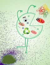
Graphical abstract

Overview of assays observing elimination of tauP301L aggregates with AUTOTAC. (A) Description of the biological working mechanism of heterobifunctional chimeric AUTOTAC degraders. (B) Schematic illustration of assays described in this paper.
Background
Targeted protein degradation (TPD) is a global protein editing tool with the potential to modulate the half-life of any proteinaceous cargoes (Chamberlain and Hamann, 2019). In addition to serving as a research tool for protein degradation and function, TPD offers an alternative—and thus attractive—means to eradicate disease-causing agents, especially those entrenched in the previously-considered undruggable proteome (Moon and Lee, 2018; Fisher and Phillips, 2018; Schapira et al., 2019; Ji et al., 2022). The various TPD platforms to date harness either the proteasome or the lysosome to facilitate the targeted elimination of a wide variety of cargoes, both within and outside the cell via distinct mechanisms, exemplified by PROteolysis-Targeting Chimera (PROTAC), Antibody-based proTAC (AbTAC), Lysosome-Targeting Chimera (LYTAC), Autophagy-Targeting Chimera (AUTAC), and AuTophagosome-TEthering Compound (ATTEC), to name just a few (Takalo et al., 2013; Thibaudeau et al., 2018; Takahashi et al., 2019; Li et al., 2019; Banik et al., 2020).
Despite the rapid development of TPD platforms with distinct mechanisms of action, the large majority of focus has been centered around a relatively few number of targets, mostly oncoproteins. Even with the emergence of autophagy/lysosome-based degraders, efficient degradation has been limited to only a handful of alternative targets—especially those responsible for neurodegeneration and proteopathies in general. Consequently, development of novel autophagy-based degraders that can target a wide range of pathological hallmark proteins of neurodegeneration and proteopathies, most notably their high-molecular weight, oligomeric, aggregated, and amyloidogenic species, still remains a pressing issue. A notable obstacle to such development is the relative lack of in vitro and in vivo methodologies to analyze in depth the degradative potency and mechanism of action of such degraders. Such difficulty is exacerbated by the fact that, unlike oncoproteins, the downstream signaling cascades of hallmark proteins of neurodegeneration/proteopathies still remain murky, which accentuates even further the importance of well-defined, precise assays and models to assess the autophagic mechanism of action and potency of an autophagy-based degrader (Peeraer et al., 2015; Lim et al., 2018).
To that end, we have recently reported the development and proof-of-concept characterization of a novel p62-targeting/activating TPD platform called AUTOphagy-TArgeting Chimera (Ji et al., 2022). AUTOTAC degraders can selectively recognize and target a broad range of cellular proteins to autophagic membranes for lysosomal degradation, through the selective interaction and activation of the archetypal autophagy cargo receptor p62/SQSTM1 via its ZZ domain. This interaction induces the conformational activation of inactive p62 into an autophagy-compatible form, which exposes PB1 and LIR domains, and facilitates p62 self-oligomerization in complex with targets and its interaction with LC3 on autophagic membranes (Cha-Molstad et al., 2015; Cha-Molstad et al., 2017; Ji et al., 2017, 2019, 2020; Zhang et al., 2018; Yoo et al., 2018; Heo et al., 2021, 2022). AUTOTAC technology was shown to efficiently degrade both oncoproteins and misfolded, aggregation-prone proteins of neurodegeneration.
In this paper, adopted from our paper describing the proof-of-concept development of the AUTOTAC platform, we describe both in vitro methods and in vivo models to not only induce pathological tau aggregation but also assess the selective degradative potency of a given TPD platform towards said aggregates and fibrils (Ji et al., 2022). To this end, we employed SH-SY5Y-hTauP301L cell lines and murine model that can utilize various fluorescent reporters, including full or split halves of the GFP protein, as well as a tandem mRFP-GFP fusion (Shin et al., 2020). Using these models, we induced the formation of detergent-insoluble, high-molecular weight aggregates of the mutant recombinant tau both in vitro and in vivo. The AUTOTAC TPD platform was shown to successfully degrade these pathological tau species in a lysosome-dependent manner. Overall, the protocol provides a systematic approach to generating pathological tau for the testing of not only AUTOTACs but also other autophagy-based degraders and small-molecule compounds in general, which may be expanded to other hallmark proteins of misfolding and aggregation-prone proteopathies and neurodegeneration.
Materials and Reagents
Part I. In vitro degradation of tauP301L aggregates
5 mL pipette aid (SPL, catalog number: 91010)
10 mL pipette aid (SPL, catalog number: 91005)
1.5 mL e-tube (Axygen, catalog number: MCT-150-C)
100 mm dish (Nest, catalog number: 704002, SPL, catalog number: 30100)
6-well plate (Nest, catalog number: 703003, SPL, catalog number: 30006)
12-well plate (Nest, catalog number: 712003, SPL, catalog number: 30012)
24-well plate (Nest, catalog number: 702002, SPL, catalog number: 30024)
Kim-wipe or paper towel (Yuhan-Kimberly, catalog number: 41112)
Polyvinylidene difluoride membrane, 0.45 μm (Millipore, catalog number: IPVH00010)
X-ray film (AGFA, catalog number: CP-BU)
Microscope slide (Marienfield, catalog number: 1000612)
Cover glass (Marienfield, catalog number: 0117520)
PBS (Biosesang, catalog number: P2007-1)
DMEM (Life Technologies, Gibco®, catalog number: 11995-065)
RPMI (Life Technologies, Gibco®, catalog number: 22400-089)
Opti-mem (Life Technologies, Gibco®, catalog number: 31984-070)
0.05% trypsin (Life Technologies, Gibco®, catalog number: 25300)
Lipofectamine 2000 (Invitrogen, catalog number: 11668019)
Sodium dodecyl sulfate (SDS) (Duchefa Biochemie, catalog number: S1377.1000)
Triple-distilled water
1 M Tris-HCl, pH 6.8 (Biosesang, catalog number: TR2016-050-68)
1.5 M Tris-HCl, pH 8.8 (Biosesang, catalog number: TR4020-050-88)
APS (Ammonium Persulfate) (Bio-Rad, catalog number: 1610700)
TEMED (Bio-Rad, catalog number: 161-0801)
2-Propanol (Sigma-Aldrich, catalog number: L53191334)
5× Laemmli sample buffer (Elpis Biotech, catalog number: EBA-1052)
Skim milk (Georgiachem, catalog number: SM2010)
SuperSignalTM West PICO PLUS (Thermo Fisher Scientific, catalog number: 34578)
PFF (Recombinant Human Tau P301L pre-formed fibrils) (NOVUS, catalog number: NBP2-76794)
Hydroxychloroquine (HCQ) (Sigma, catalog number: A25547)
Okadaic acid (Enzo, catalog number: ALX-350-003-C100)
HEPES (Duchefa Biochemie, catalog number: H1504.011)
PierceTM protein transfection reagent (PJ) (Thermo Fisher Scientific, catalog number: 89850)
NaCl (Duchefa Biochemie, catalog number: S0520.5000)
RIPA (Cell nest, catalog number: CNR001-0100)
PierceTM BCA protein assay kit (Thermo Fisher Scientific, catalog number: 23225)
4× LDS sample buffer non-reducing (Thermo Fisher Scientific, 84788)
PLL solution (Sigma-Aldrich, catalog number: P4707)
Albumin (Biosesang, catalog number: AC1025-100-00)
Foil (SAM JIN FOIL, 30 cm × 30 m)
Triton X-100 (Sigma-Aldrich, catalog number: T9284)
Antifade-mounting media with DAPI (Vectashield, catalog number: H-1500)
Fractionation buffer (see Recipes)
SDS-detergent lysis buffer (see Recipes)
SDS-PAGE gel (see Recipes)
Part II. In vivo elimination of tauP301L neurofibrillary tangles
Injection syringes (Becton Dickinson Medical, catalog number: 1258696)
Dissection stainless steel tray (any brand)
Pins or tape (any brand)
Butterfly needles (Biosigma, catalog number: BSS250)
25- or 27-gauge infusion needle (KOREAVACCINE, catalog number: K201)
15 mL conical tube (Tarsons, catalog number: 546021)
1.5 mL e-tube (Axygen, catalog number: MCT-150-C)
24-well plate (Nest, catalog number: 702002, SPL, catalog number: 30024)
Kim-wipe or paper tower (Yuhan-Kimberly, catalog number: 41112)
Foil (SAM JIN FOIL, 30 cm × 30 m)
Microscope slide (Marienfield, catalog number: 1000612)
PBS (Biosesang, catalog number: P2007-1)
Avertin (Sigma-Aldrich, catalog number: T48402)
0.9% saline (Sigma-Aldrich, catalog number: 08059)
RIPA (Cell nest, catalog number: CNR001-0100)
Protease inhibitor (Abbkine, catalog number: BMP1001)
Phosphatase inhibitor (Sigma-Aldrich, catalog number: P0001)
Sucrose (Sigma-Aldrich, catalog number: S0389)
Sodium dodecyl sulfate (SDS) (Duchefa Biochemie, catalog number: S1377.1000)
Triple-distilled water (DIW)
4% paraformaldehyde (Biosesang, catalog number: pc2031-100-00)
Optional cutting temperature (OCT) compound (Sakura Tissue-Tek, catalog number: 4583)
Sodium azide (Thermo Fisher Scientific, catalog number: J21610-22)
Sudan black B (Sigma-Aldrich, catalog number: 199664)
Hoechst (Abcam, ab228550)
EtOH (DUKSAN, catalog number: 64-17-5)
0.01% PBST (see Recipes)
Blocking solution (see Recipes)
Injection drug (see Recipes)
Primary antibodies
Rabbit polyclonal anti-LC3 (Sigma-Aldrich, catalog number: L7543, 1:10,000 diluted in PBST solution)
Rabbit polyclonal anti-GAPDH (Bio World, catalog number: AP0063, 1:10,000 diluted in PBST solution)
Mouse monoclonal anti-Tau5 (Invitrogen, catalog number: AHB0042, 1:5,000 diluted in PBST solution)
Rabbit polyclonal anti-p-Tau (Invitrogen, catalog number: 44–752G, 1:5,000 diluted in PBST solution)
AT8 antibody (Abcam, catalog number: ab210703, 1:200 diluted in the blocking solution)
Secondary antibodies
Anti-rabbit IgG-HRP (Cell Signaling, catalog number: 7074, 1:10,000 diluted in PBST solution)
Anti-mouse IgG-HRP (Cell Signaling, catalog number: 7076, 1:10,000 diluted in PBST solution)
Cell
HeLa (ATCC, catalog number: CCL-2): the first immortal human cells to be grown in culture and the basis for countless scientific discoveries
SH-SY5Y-tauP301L-GFP (Innoprot): SH-SY5Y stable cells tagged with green fluorescence and expressing tau mutant [0N4R(P301L mutant)]
Mouse
TauP301L-BiFC transgenic mouse (Shin et al., 2020): the mouse model that has two non-fluorescent compartments (hTauP301L-VN173 and hTauP301L-VC155) turning on Venus fluorescence protein upon tau assembly when they are fused with TauP301L.
Plasmid
mRFP-GFP-hTauP301L plasmid [mRFP-GFP-LC3 plasmid (Cha-Molstad et al., 2016; Dalby et al., 2004) is modified with hTauP301L]
Equipment
Part I. In vitro degradation of tauP301L aggregates
Tabletop centrifuge (Eppendorf, model/catalog number: 5424R)
Sonicator (SONICS vibra cel)
Centrifuge (Hanil fieta 4)
37°C incubator (Hera cell vios 250i CO2 incubator)
Laser scanning confocal microscope (Zeiss 510 Meta)
Rocker, CR300 (Finepcr)
Part II. In vivo elimination of tauP301L neurofibrillary tangles
Dissecting forceps (JEUNG DO BIO&PLANT CO., catalog number: SD-S-04PK)
Tissue scissors (JEUNG DO BIO&PLANT CO., catalog number: S-49-16-SPK, S-54-10-SPK)
Vascular clamp (Surtex Instruments)
Micro spatula (Merck, catalog number: Z243213)
Peristaltic pump (Ismatec., catalog number: ISM834)
7 mL Dounce tissue grinder (Wheaton, catalog number: 35742)
Tissue grinder motor (Bel-Art, KA., catalog number: UB32-50)
Polypropylene pestle (Bel-Art, BA., catalog number: 19923-0001)
Rotor (FINEPCR, AG)
Tabletop centrifuge (Eppendorf, catalog number: 5424R)
-80°C deep freezer (Thermo Fisher Scientific, catalog number: TDE)
Cryostat (Leica, catalog number: CM3050S)
Cryomolds (Sakura Tissue-Tek, catalog number: 25608-922)
Paintbrush (AnB, catalog number: P000EACE)
ZEISS Axio Scan.Z1 (Carl Zeiss, Germany)
Software
LMS image software (Zeiss LSM Image Browser (ver. 4.2.0.121))
ImageJ (NIH, Bethesda)
Prism 6 software (GraphPad)
Procedure
文章信息
版权信息
© 2023 The Authors; exclusive licensee Bio-protocol LLC.
如何引用
Lee, M. J., Kim, S. B., Kim, H. Y., Lee, S. J., Lee, J. S., Kwon, Y. T. and Ji, C. H. (2023). Methods to Detect AUTOphagy-Targeting Chimera (AUTOTAC)-mediated Targeted Protein Degradation in Tauopathies. Bio-protocol 13(2): e4594. DOI: 10.21769/BioProtoc.4594.
分类
药物发现 > 药物筛选
神经科学 > 神经系统疾病 > 细胞机制
细胞生物学 > 基于细胞的分析方法 > 自噬活性
您对这篇实验方法有问题吗?
在此处发布您的问题,我们将邀请本文作者来回答。同时,我们会将您的问题发布到Bio-protocol Exchange,以便寻求社区成员的帮助。