- EN - English
- CN - 中文
Fluorescence Time-lapse Imaging of Entosis Using Tetramethylrhodamine Methyl Ester Staining
使用四甲基罗丹明甲酯染色对内吞死亡进行荧光延时成像
发布: 2022年12月05日第12卷第23期 DOI: 10.21769/BioProtoc.4564 浏览次数: 1885
评审: Ralph Thomas BoettcherRakesh BamOlga Kopach
Abstract
Entosis is a process where a living cell launches an invasion into another living cell’s cytoplasm. These inner cells can survive inside outer cells for a long period of time, can undergo cell division, or can be released. However, the fate of most inner cells is lysosomal degradation by entotic cell death. Entosis can be detected by imaging a combination of membrane, cytoplasmic, nuclear, and lysosomal staining in the cells. Here, we provide a protocol for detecting entosis events and measuring the kinetics of entotic cell death by time-lapse imaging using tetramethylrhodamine methyl ester (TMRM) staining.
Keywords: Entosis (内吞死亡)
Background
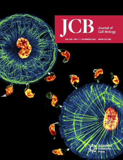
Entosis is a form of cell–cell interaction in which a living cell (inner cell) orchestrates its invasion into another cell (outer cell) by a Rho/ROCK signaling–dependent mechanism (Overholtzer et al., 2007). Entosis results in the formation of a unique morphological microscopic structure, also known as cell-in-cell structure or bird’s eye structure, which is characterized by evidence of an inner cell, a visible entotic vacuole between inner and outer cells, and an outer cell with a crescent-shaped nucleus as it is pushed towards the periphery (Fais and Overholtzer, 2018). Entosis spontaneously—but rarely—occurs in cultured cells in vitro; nevertheless, it has important consequences for both inner and outer cells. From one perspective, entosis seems to be an assisted-suicide mechanism for the inner cells, as the vast majority of these undergo lysosomal degradation by a mechanism known as entotic cell death (Florey et al., 2011). However, inner cells can also survive and be released, and even complete full cell division inside outer cells. Intriguingly, outer cells also benefit from this invasion as they gain survival advantage under stress conditions (Hamann et al., 2017) or become resistant to apoptosis (Bozkurt et al., 2021). Entosis-like structures are more frequently observed in cancers compared to normal tissues, and the presence of these structures is associated with poor outcome and disease recurrence in cancer (Mackay et al., 2018; Bozkurt et al., 2021).
Quantification of entosis relies on morphological detection of the events using microscopy images. Currently, it is not possible to automatically quantify entosis events when both inner and outer cells are alive. Thus, events are first detected with the aid of nuclear, cytoplasmic, and/or membrane staining, and then manually quantified (Overholtzer et al., 2007). When inner cells undergo entotic cell death, however, quantifying entosis events is relatively easy due to lysosomal acidification of inner cells. Lysosomal marker Lamp1 or autophagic marker LC3 can be fluorescently expressed in the cells to quantify the dynamics of entotic cell death (Florey et al., 2011; Overholtzer et al., 2007). However, overexpression of artificially produced proteins might also affect the rate of entosis events. Probably the most practical method to label entotic cell death is using lysosomal dyes such as LysoTracker, which freely passes the cell membrane and is sequestered inside acidic organelles. Alternatively, fluorogenic cathepsin substrates or acridine orange have been used to detect entosis and entotic cell death (Overholtzer et al., 2007; Garanina et al., 2017).
Recently, we reported a novel alternative approach for detecting entosis events and measuring the dynamics of entotic cell death by using tetramethylrhodamine methyl ester (TMRM) (Bozkurt et al., 2021). This cationic cell-permeable dye is sequestered by active mitochondria and has been widely used to analyze real-time changes in mitochondrial events such as loss of mitochondrial membrane potential during apoptosis as well as fusion/fission (Dußmann et al., 2003; Cho et al., 2019). Here, we provide a detailed protocol for performing time-lapse microscopy in any cell line to detect entosis events and entotic cell death by using TMRM staining. Our approach allows simultaneous detection of mitochondrial and entotic events as well as quantification of the real-time kinetics in living cells at the single-cell level.
Materials and Reagents
WillCo-dish® 12 mm glass bottom dishes (WillCo Wells B.V., catalog number: HBST-3512)
Cell culture flask, T-75, surface: standard, filter cap (Sarstedt, catalog number: 83.3911.002)
Living cells (in this protocol, HCT116-Venus cells (Bozkurt et al., 2021) were used; however, this method is applicable to any living cell)
Fetal bovine serum (FBS) (Sigma-Aldrich, catalog number: F7525), store at -20 °C.
L-glutamine (Sigma-Aldrich, catalog number: G7513), store at -20 °C
Mineral oil, suitable for mouse embryo cell culture (Sigma-Aldrich, catalog number: M5310), store at room temperature
Penicillin–streptomycin (Sigma-Aldrich, catalog number: P0781), store at -20 °C
Roswell Park Memorial Institute 1640 medium (RPMI medium) (Sigma-Aldrich, catalog number: R0883), store at 4 °C
Tetramethylrhodamine, methyl ester, perchlorate (TMRM) (Thermo Fisher Scientific, catalog number: T668), store at 4 °C
Equipment
Inverted confocal laser scanning microscope (Carl Zeiss Ltd, LSM 710) equipped with a 40×/1.3 NA plan apochromat oil immersion objective, a microscope incubator chamber (37 °C with 5% CO2), and a motorized stage
Cell culture hood (Heraeus, HERAsafe, Type HS12)
Centrifuge (Eppendorf, 5810, catalog number: 5810000060)
Water bath (GFL Type 1004)
Humidified CO2 incubator (New Brunswick/Eppendorf, Galaxy 170S)
Software
ZEN 2009 version 6,0,0,303 configuration 5, with MTS 2009-2010 version 32.007
Fiji/ImageJ (National Institutes of Health, https://imagej.nih.gov/ij/)
PlotTwist, a web app for plotting continuous data (https://huygens.science.uva.nl/PlotTwist/) (Goedhart, 2020)
Procedure
文章信息
版权信息
© 2022 The Authors; exclusive licensee Bio-protocol LLC.
如何引用
Readers should cite both the Bio-protocol article and the original research article where this protocol was used:
- Bozkurt, E., Düssmann, H. and Prehn, J. H. M. (2022). Fluorescence Time-lapse Imaging of Entosis Using Tetramethylrhodamine Methyl Ester Staining. Bio-protocol 12(23): e4564. DOI: 10.21769/BioProtoc.4564.
- Bozkurt, E., Düssmann, H., Salvucci, M., Cavanagh, B. L., Van Schaeybroeck, S., Longley, D. B., Martin, S. J. and Prehn, J. H. (2021). TRAIL signaling promotes entosis in colorectal cancer. J Cell Biol 220(11): e202010030.
分类
癌症生物学 > 细胞死亡 > 细胞生物学试验
细胞生物学 > 细胞分离和培养 > 单层培养
细胞生物学 > 细胞成像 > 活细胞成像
您对这篇实验方法有问题吗?
在此处发布您的问题,我们将邀请本文作者来回答。同时,我们会将您的问题发布到Bio-protocol Exchange,以便寻求社区成员的帮助。
Share
Bluesky
X
Copy link


