

Analysis of Caenorhabditis elegans Aging-related Neurodegeneration in Chemosensory Neurons
秀丽隐杆线虫化学感觉神经元衰老相关神经退行性变分析

发布: 2022年07月20日第12卷第14期 DOI: 10.21769/BioProtoc.4473 浏览次数: 2658
评审: Anonymous reviewer(s)
参见作者原研究论文
The authors used this protocol in:
2020


Abstract
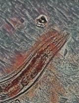
Aging and neuronal deterioration constitute important risk factors for the development of neuronal-related diseases, such as different dementia. The nematode Caenorhabditis elegans has emerged as a popular model system for studying neurodegeneration diseases, due to its complete neuronal connectivity map. DiI is a red fluorescent dye that can fill the worm amphid neurons and enables the visualization of their neurodegeneration over time. This protocol provides an efficient, fast, and safe method to stain worm amphid neurons to highlight the chemosensory structures of live nematodes.
Keywords: DiI (细胞膜红色荧光探针)Background
Neurodegenerative diseases share a common predisposing factor, the aging of the brain. So, a greater understanding of the normal aging brain may be necessary before we can fully understand the causes of pathological aging and cognitive decline (Yankner et al., 2008).
The existence of a complete neuronal connectivity map and genetic tractability of C. elegans make this animal model useful for studying human neurological diseases. Analysis of multiple genetic databases shows that a considerable number of human genes associated with different diseases have a significant homology to C. elegans genes, and the genetic tools available for this nematode have allowed the construction of predictive models for studying the molecular mechanism of these diseases (Alexander et al., 2014; Griffin et al., 2017).
In the aging C. elegans nervous system, synapses appear physically less robust than in younger animals. The average synapse in an elderly animal is smaller in its presynaptic density and includes fewer synaptic vesicles compared to young adults; this is similar to what occurs in the human brain, where loss of synaptic integrity contributes to its deterioration and dysfunction. Away from the synapse, typical changes in aging neurons include reduced axon caliber, poor axonal transport, smaller nuclei, and reduced cytoplasmic volume. In addition, part of the loss of mobility has an origin in defects of the nervous system (Toth et al., 2012). In recent years, numerous protocols have emerged to facilitate the visualization of C. elegans neurons, taking advantage of the transparent nature of this nematode. DiI is a long-chain dialkylcarbocyanine dye that uniformly labels neurons via lateral diffusion in the plasma membrane (Godement et al., 1987; Hofmann and Bleckmann, 1999). Neurons can be labeled either retrogradely or during dissociation. Some of the labeled membrane gradually becomes internalized and retains its fluorescence, allowing identification of cells for several weeks. DiI specifically labels amphid (ADL, ASH, ASI, ASJ, ASK, and AWB), and phasmid (PHA and PHB) IL1 and IL2 neurons, as well as IL sheath and socket cells to highlight the chemosensory structures of live nematodes (Wormatlas.org [Reference 12]). These dyes do not appear to affect the survival, development, or basic physiological properties of neurons, and do not spread detectably from labeled to unlabeled neurons. It seems likely that cells become retrogradely labeled mainly by lateral diffusion of dye in the plane of the membrane (Honig and Hume, 1986). Labeling with carbocyanine dyes has already allowed several exciting advances in developmental neurobiology (Honig and Hume, 1989). The aim of this protocol is to provide a modified technique for Dil staining, to allow the study of neurodegeneration in dementia disease models of C. elegans.
Materials and Reagents
Petri dishes 60 × 15 mm 500/cs (Fisher Scientific, catalog number: FB0875713A)
Pipette tips 2–200 µL Eppendorf® epT.I.P.S. (Eppendorf, catalog number: 022492039)
Pipette tips 50–1,000 µL Eppendorf® epT.I.P.S. (Eppendorf, catalog number: 022492055)
Eppendorf® Safe-Lock 1.5 mL microcentrifuge tubes (Eppendorf, catalog number: 022363204)
Corning® 15 mL centrifuge tubes (Corning, catalog number: 430791)
Glass media bottles 200 mL (Fisher Scientific, catalog number: FB800250)
Glass slide (25 × 75 × 0.9mm) and coverslip (22 × 22mm) (Sigma-Aldrich, catalog number: CLS294875X25-72EA; C9802-1PAK)
C. elegans strains (Caenorhabditis Genetics Center: N2 Bristol)
Escherichia coli strain OP50-1 (University of Minnesota, C. elegans Genetics Center, MN)
Bacillus subtilis strain NCIB3610 (Bacillus Genetic Stock Center, catalog numbers: 3A1 and 1A96)
1,1’-dioctadecyl-3,3,3’,3’,-tetramethylindocarbocyanine perchlorate (DiI, Sigma-Aldrich, catalog number: 468495)
Magnesium sulfate heptahydrate (MgSO4·7H2O) (Sigma-Aldrich, catalog number: M1880)
Potassium phosphate monobasic (KH2PO4) (Sigma-Aldrich, catalog number: P5655)
Potassium phosphate dibasic (K2HPO4) (Sigma-Aldrich, catalog number: P2222)
Sodium chloride (NaCl) (Sigma-Aldrich, catalog number: S7653)
Sodium azide (Sigma-Aldrich, catalog number: S8032)
NaClO Hypochlorite (Sigma-Aldrich, catalog number: 13440)
Sodium hydroxide (NaOH) (Sigma-Aldrich, catalog number: S8045)
Bacto peptone (BD, BactoTM, catalog number: 211677)
Agar (Sigma-Aldrich, catalog number: A1296)
Cholesterol in absolute ethanol Cholesterol (Sigma-Aldrich, catalog number: C8667)
100% ethanol (Sigma-Aldrich, catalog number: E7023)
Calcium chloride dihydrate (CaCl2·2H2O) (Sigma-Aldrich, catalog number: C3881)
Luria broth (Sigma-Aldrich, catalog number: L3522)
M9 buffer (see Recipes)
Nematode growth medium (NGM) agar (see Recipes)
Potassium phosphate buffer (see Recipes)
1 M MgSO4 (see Recipes) 5 mg/mL cholesterol (see Recipes)
1 M CaCl2 (see Recipes)
LB media (see Recipes)
Phosphate buffer (see Recipes)
1 N NaOH (see Recipes)
Bleaching solution (see Recipes)
DiI stock solution (see Recipes)
Equipment
Fluorescence microscope Zeiss (Carl Zeiss, Serial number: 3108012846)
Shaker (Axyos Technologies, catalog number: S04036.tu)
Autoclave (Tuttnauerusa, model: 6690)
Worm pick. Worm picks can either be purchased (Genesee Scientific, catalog number: 59-AWP) or made in the lab as described in Wollenberg et al. (2013)
Centrifuge (Eppendorf, model: 5430)
Tabletop centrifuge (Eppendorf, model: 5424)
Bunsen burner (Humbolt, catalog number: H-5870)
Procedure
文章信息
版权信息
© 2022 The Authors; exclusive licensee Bio-protocol LLC.
如何引用
Crespo, C. and Grau, R. (2022). Analysis of Caenorhabditis elegans Aging-related Neurodegeneration in Chemosensory Neurons. Bio-protocol 12(14): e4473. DOI: 10.21769/BioProtoc.4473.
分类
神经科学 > 神经解剖学和神经环路 > 动物模型
发育生物学 > 形态建成
生物科学 > 生物技术 > 微生物技术
您对这篇实验方法有问题吗?
在此处发布您的问题,我们将邀请本文作者来回答。同时,我们会将您的问题发布到Bio-protocol Exchange,以便寻求社区成员的帮助。