

Immunohistochemistry of Immune Cells and Cells Bound to in vivo Administered Antibodies in Liver, Lung, Pancreas, and Colon of B6/lpr Mice
B6/lpr小鼠肝脏、肺、胰腺和结肠中免疫细胞和与体内施用的抗体结合的细胞的免疫组织化学
发布: 2022年07月20日第12卷第14期 DOI: 10.21769/BioProtoc.4468 浏览次数: 3608
评审: Xiaoyi ZhengXiaoyu LiuEmilie Besnard
参见作者原研究论文
The authors used this protocol in:

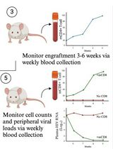
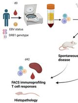
Feb 2021
Advertisement


Abstract
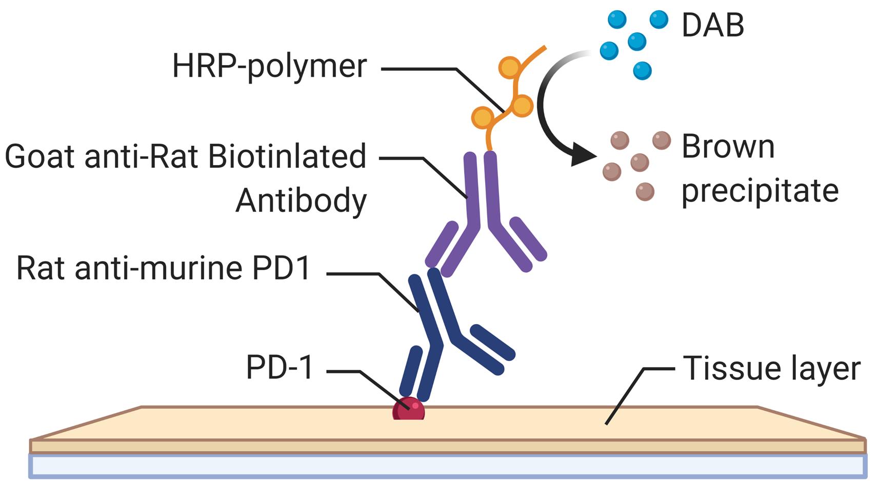
Employing a novel mouse model of immune related adverse events (irAEs) induced by combination of anti-PD1 and anti-CTLA-4 antibodies, we visualized immune infiltration into the liver, lung, pancreas, and colon. Here, we describe the avidin-biotin conjugate (ABC) method used to stain T cells (CD4 and CD8), B cells (CD19), macrophages (F4/80), and cells bound by the in vivo administered rat anti-mouse antibodies for chromogenic immunohistochemistry (IHC). Using a biotinylated goat anti-rat antibody, we detected the localization of cells bound to the in vivo antibodies for PD-1 and CTLA-4. IHC has advantages over other techniques, namely antibody availability, resistance to photobleaching, and greater sensitivity. Additionally, detection and localization of in vivo antibodies can be used in mice models to infer their therapeutic efficacy, stability, and function.
Graphical abstract:
Background
The last decade has seen the approval of several immune checkpoint blockade (ICB) antibodies in various cancers, which has improved patient survival. However, patients that respond to these ICB develop toxicities known as irAEs. We have previously developed a novel mouse model of irAEs in B6/lpr mice, that developed multiorgan toxicities with immune infiltration, when given a combination of anti-PD-1 and anti-CTLA-4 (Adam et al., 2021). Mice were injected with 200 μg of anti-PD1 (BioXCell BE0146), and 100 μg of CTLA-4 (BioXcell BE0131) antibodies intraperitoneally twice a week for 6 weeks. Mice were sacrificed at endpoint, and tissue collected for IHC, to assess the cellular landscape. The injected ICB antibodies derived from rat were visualized in the tissue using a goat anti-rat that recognized the fragment crystallizable (Fc) portion of the IgG rat antibody. This method of detection could be used to evaluate ICB antibodies for toxicity in mice models. Although there are numerous other techniques for visualization or detection of antibodies (de Boer et al., 2015; Mestel, 2017; Knight et al., 2019; Wen et al., 2021), the limitations are the access to large and expensive microscopes, CT scanner, or MRI, which may be inaccessible to many laboratories. Our method is advantageous due to a relatively low expense, accessible selection of conjugated antibodies, and flexibility with IHC kits.
Materials and Reagents
Tissue Tek Slide Staining Dish (Fisher, catalog number: NC0731403)
Slide Holder (Fisher, catalog number: NC9697666
10 mL pre-filled flush syringe saline (NaCl 0.9%) (BD, catalog number: 306575)
25G × ¾’’ blood collection set (BD, catalog number: 367285)
FisherbrandTM ColorFrostTM Plus Microscope Slides (Fisher, catalog number: 12-550-17)
FisherbrandTM Standard Disposable Transfer Pipettes, Nongraduated (Fisher, catalog number: 13-711-7M)
Goat anti-rat IgG antibody (H+L), biotinylated (Vector, catalog number: BA-9400)
Goat anti-rabbit IgG antibody (H+L), biotinylated (Vector, catalog number: BA-1000)
VECTASTAIN(r) Elite ABC-HRP Kit, peroxidase (Standard) (Vector, catalog number: PK-6100)
Dako liquid 3,3'-diaminobenzidine (DAB) and substrate (DAKO, catalog number: K3468)
CD4 (D7D2Z) (CST, catalog number: 25229)
CD8a (D4W2Z) (CST, catalog number: 98941)
CD19 (D4V4B) (CST, catalog number: 90176)
F4/80 (BM8) (Invitrogen, catalog number: 144801)
Xylene (Sigma, catalog number: 534056)
10× Citrate buffer pH 6.0 (Novus Biologicals, catalog number: NB900-62075)
Triton X-100 (Sigma, catalog number: X100)
Ethanol (Fisher Bioreagents, catalog number: BP2818100)
Hydrogen peroxide (BioRad, catalog number: BUF017B)
Hematoxylin counterstain (Vector, catalog number: H-3401-500)
Proteinase K (abcam, catalog number: ab64220)
10× PBS (Phosphate Buffered Saline) (Thermo Scientific, catalog number: AAJ75889K8)
Normal goat serum blocking solution (Vector, catalog number: S-1000-20)
FisherbrandTM SuperfrostTM ExcellTM Microscope Slides (Fisher, catalog number: 22-037-247)
Fisher ChemicalTM PermountTM Mounting Medium (Fisher, catalog number: SP15-100)
Dako Antibody Diluent (Agilent, catalog number: S0809)
Deionized water
B6/lpr (Jackson Laboratory, catalog number: 000482)
10% Neutral Buffered Formalin (Fisher, catalog number: 22-110-869)
Globe Scientific Rectangular Cover Glass size 24 × 50 mm, No.1 (Fisher, catalog number: 22-170-388)
Tween 20 (Sigma, catalog number: P1379)
PBS (Phosphate Buffered Saline) and PBS-T (Tween) (see Recipes)
10 mM Sodium Citrate (see Recipes)
Bluing solution (see Recipes)
Equipment
Forceps
Microtome (any supplier)
Microwave (any supplier)
Pressure cooker (any supplier)
Leica SCN400 scanner
Fume hood (any supplier)
Light microscope with 20× and 40× lenses
Water bath (ISOtemp205)
Fisher Scientific Isotemp 650D incubator oven
Software
AperioImage Scope (Leica, https://www.leicabiosystems.com/us/digital-pathology/manage/aperio-imagescope)
Procedure
文章信息
版权信息
© 2022 The Authors; exclusive licensee Bio-protocol LLC.
如何引用
Adam, K. and Mor, A. (2022). Immunohistochemistry of Immune Cells and Cells Bound to in vivo Administered Antibodies in Liver, Lung, Pancreas, and Colon of B6/lpr Mice. Bio-protocol 12(14): e4468. DOI: 10.21769/BioProtoc.4468.
分类
免疫学 > 动物模型 > 小鼠
免疫学 > 免疫细胞染色 > 免疫检测
细胞生物学 > 细胞染色 > 蛋白质
您对这篇实验方法有问题吗?
在此处发布您的问题,我们将邀请本文作者来回答。同时,我们会将您的问题发布到Bio-protocol Exchange,以便寻求社区成员的帮助。