

Quantitative and Anatomical Imaging of Human Skin by Noninvasive Photoacoustic Dermoscopy
无创光声皮肤镜对人体皮肤的定量和解剖成像
发布: 2022年04月05日第12卷第7期 DOI: 10.21769/BioProtoc.4372 浏览次数: 2301
评审: Pilar Villacampa AlcubierreXinyi WangMatthias Rieckher
参见作者原研究论文
The authors used this protocol in:
Jan 2021
Advertisement


Abstract
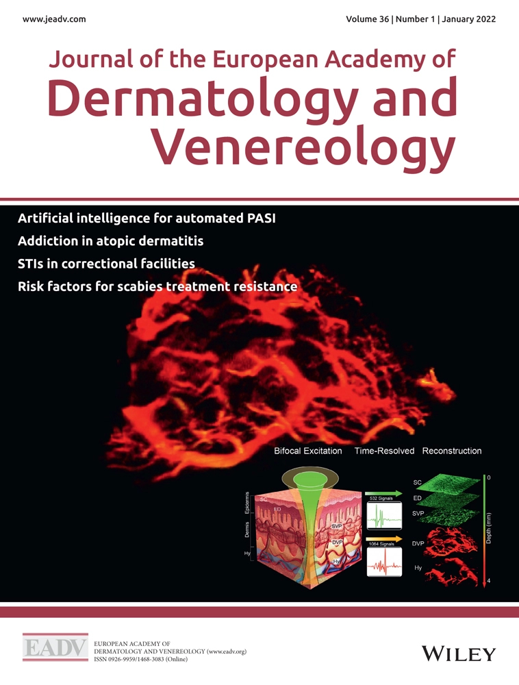
Imaging plays a vital role in the diagnosis and treatment of skin diseases. However, pure optical imaging technique is limited to the visualization of superficial skin tissues. Ultrasonic imaging technique can detect deep tissues, but it lacks detailed information on microscopic pathological structures. Photoacoustic imaging is an advanced technology that bridges the spatial-resolution gap between optical and ultrasonic techniques, by the modes of optical excitation and acoustic detection. Photoacoustic dermoscopy (PAD), based on photoacoustic technology, can noninvasively obtain high-resolution anatomical structures by endogenous absorbers, such as melanin, hemoglobin, lipids, etc. In the past years, PAD has gradually been developed in clinical dermatology for the diagnosis of melanoma, psoriasis, port-wine stains, dermatitis, skin grafting, and testing the efficacy of cosmetics. This protocol provides detailed procedures for PAD construction, including component selection, equipment setup, and system calibration. A step-by-step guide for human skin imaging is provided as an example application. Image reconstruction and troubleshooting procedures are also elaborated. PAD offers the 3D volumetric images of human skin, and quantitatively analyzes the vascular morphology in the dermis. The protocol will provide clinicians with standardized and reasonable guidance in dermatological imaging.
Keywords: Biomedical imaging (生物医学成像)Background
The skin is the largest organ of the human body. The state of the skin is closely related to human health. Imaging technology plays an important role in dermatology. Conventional pure optical dermatoscopes are limited by imaging depth and can only visualize epidermal morphology. Ultrasound imaging can visualize the entire skin structure with deep penetration, but its spatial resolution and ability to visualize microvessels are relatively poor. Photoacoustic imaging (PAI), based on the photoacoustic effect, is a hybrid biomedical imaging technology, which combines high-contrast optical imaging and deep penetration ultrasound imaging (Wang and Hu, 2012). As a meritorious application mode of PAI, photoacoustic dermoscopy (PAD) provides researchers and clinicians with a new way of noninvasively observing deep skin structures (Xu et al., 2016; Ma et al., 2018, 2020, 2021; Wang et al., 2022a, 2022b). PAD overcomes the strong photon scattering caused by the skin tissue’s multilayered structures and complex heterogeneous medium, and achieves full-thickness imaging of human skin with high resolution (Wang et al., 2022b). Differently configured PADs allow spatial resolution to be scaled to the desired skin imaging depth, while maintaining a high depth-to-resolution ratio. By depicting the fine anatomical structures from epidermis to subcutaneous tissue, PAD can be used to diagnose abnormal pigmentation, and vascular malformation. PAD is gradually proposed in clinical dermatology for the diagnosis of melanoma, psoriasis, port-wine stains, dermatitis, skin grafting, and for the assessment of the efficacy of cosmetics (Aguirre et al., 2017; Tsuge et al., 2020; Wang et al., 2022a). In clinical diagnosis and treatment, quantitative information on the distribution, depth, and diameter of the melanin and vessels can be obtained to determine optimum treatment parameters for individual patients. Currently, the limitation of PAD lies in the fact that coupling gel is required to transmit photoacoustic signals, which is not conducive to imaging skin ulcers. In the future, non-contact photoacoustic detection technology could make up for this deficiency. This protocol is dedicated to the design and debugging of PAD for human skin imaging. Overall, this protocol will promote preclinical and clinical applications of PAD in dermatology and plastic surgery.
Materials
Polyethylene membrane (He Jian Jie Li Plastic Products Co., Ltd., membrane thickness: 10 μm)
Ultrasound gel (GUANGGONG PAI, medical ultrasound gel)
Deionized water
Equipment
FiberPort (Thorlab, model: PAF-X-7-A)
Single-mode fiber (Thorlab, model: P1-460B-FC-5)
Collimator (Thorlab, model: F240FC-532)
Objective lens (Daheng Optics, model: GCO-2101)
Ultrasonic transducer (Doppler Electronic Technology, model: Foused ϕ8-3)
Amplifier (RF Bay, model: LNA-650)
Data acquisition card (Spectrum, model: M3i.3221)
Graphics Processing Unit (NVIDIA, model: GeForce GTX 1060)
X-Y translation stage (Jiancheng Optics, model: LS2-25T)
Motor driver (Jiancheng Optics, model: LS2-MD)
Robot arm (Ergotron, model: 826-842)
Field Programmable Gate Array (Altera, model: Cyclone IV)
Coupling cup (Shenzhen WeNext Technology, material: nylon)
Coaxial cable (Talent RG cable, model: RG58/U)
SubMiniature version A (SMA) connector (Rebes, model: 50 Ω)
Laser goggles (Daheng Optics, model: GCAQ-YAD)
Copper foil tape (Shenzhen Sanwangda Electronic Materials, model:3M1181)
Buckle-type magnetic ring (Jia Lin Magnetic Industry, model: SCRC 35B)
532-nm pulsed laser (Laser-export, model: DTL-314QT)
DC power supply (LONG WEI, model: PS-1502D)
Laser power meter (Physcience Opto-electronics, model: LPE-1)
Digital multimeter (Fluke, model: 115C)
Software
LabVIEW 2016 (National Instruments, https://www.ni.com/zh-cn.html)
MATLAB2020a (MathWorks, https://ww2.mathworks.cn/)
ImageJ (National Institutes of Health software, https://imagej.nih.gov/ij/)
Procedure
文章信息
版权信息
© 2022 The Authors; exclusive licensee Bio-protocol LLC.
如何引用
Wang, Z., Yang, F., Zhang, W. and Yang, S. (2022). Quantitative and Anatomical Imaging of Human Skin by Noninvasive Photoacoustic Dermoscopy. Bio-protocol 12(7): e4372. DOI: 10.21769/BioProtoc.4372.
分类
生物工程 > 生物医学工程
生物物理学 > 显微技术
生物科学
您对这篇实验方法有问题吗?
在此处发布您的问题,我们将邀请本文作者来回答。同时,我们会将您的问题发布到Bio-protocol Exchange,以便寻求社区成员的帮助。