

Reconstruction of Human AML Using Functionally and Immunophenotypically Defined Human Haematopoietic Stem and Progenitor Cells as Targeted Populations
使用功能和免疫表型定义的人类造血干细胞和祖细胞重建人类急性髓细胞白血病模型
发布: 2021年12月20日第11卷第24期 DOI: 10.21769/BioProtoc.4262 浏览次数: 4440
评审: Alak MannaShyam SrivatsNavnita Dutta
参见作者原研究论文
The authors used this protocol in:
Feb 2021
Advertisement


Abstract
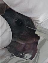
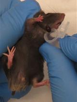
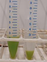
Acute myeloid leukaemia (AML) is a highly heterogenous blood cancer, in which the expansion of aberrant myeloid blood cells interferes with the generation and function of normal blood cells. Although key driver mutations and their associated inhibitors have been identified in the last decade, they have not been fully translated into better survival rates for AML patients, which remain dismal. In addition to DNA mutation, studies in mouse models strongly suggest that the cell of origin, where the driver mutation (such as MLL fusions) occurs, emerges as an additional factor that determines the treatment outcome in AML. To investigate its functional relevance in human disease, we have recently reported that AML driven by MLL fusions can transform immunophenotypically and functionally distinctive human hematopoietic stem cells (HSCs) or myeloid progenitors resulting in immunophenotypically indistinguishable human AML. Intriguingly, these cells display differential treatment sensitivities to current treatments, attesting the cell of origin as an important determinant governing treatment outcome for AML. To further facilitate this line of investigation, here we describe a comprehensive disease modelling protocol using human primary haematopoietic cells, which covers all the key steps, from the isolation of immunophenotypically defined human primary haematopoietic stem and progenitor populations, to oncogene transfer via viral transduction, the in vitro liquid culture assay, and finally the xenotransplantation into immunocompromised mice.
Background
Acute myeloid leukaemia (AML) is a highly heterogenous blood cancer driven by diverse mutations and distinctive cell populations, which is generally associated with poor prognosis in particular for older patients aged over 60. Recent research efforts have not only brought to light key AML driver mutations but also greatly enhanced our mechanistic understanding of how these mutations transform normal blood cells into leukaemia cells (Zeisig et al., 2012). Despite this progress, the survival rates of most AML patients have only marginally improved and remain dismal (Shallis et al., 2019).
Disease modelling is a powerful tool to study cancer biology, as it allows the researchers to readily access bona fide diseased cells for subsequent cellular and molecular studies. Mouse models have been particularly instrumental given the easy accessibility and well-established protocols for genetic manipulation, in vitro, and in vivo propagation of the cells. They have provided important insights into the biology of AML cells transformed by key driver mutations including MLL fusions and revealed promising novel therapeutic targets (Almosailleakh and Schwaller, 2019; Zeisig and So, 2021). Murine disease models also led to the identification of candidate leukaemia cell of origins and their potential roles in influencing treatment responses (Krivtsov et al., 2013; Stavropoulou et al., 2016; Siriboonpiputtana et al., 2017). While human and mouse cells share significant similarities, there are also clear differences including their contrasting transformation requirements, telomere lengths, and the non-coding regulatory genomes. As compared with mouse cell models, reconstruction of human AML using human primary cells has significantly laged behind (Barabé et al., 2007; Horton et al., 2013; Wunderlich et al., 2013). Until recently, it was not clear if human AML can also originate from multiple cells of origin, which may have different biology and treatment responses. Using immunophenotypically and functionally defined hematopoietic populations isolated from umbilical cord blood as the targeted cells to reconstruct MLL-AML, we have shown that human HSCs and common myeloid progenitors (CMPs) can be the cellular origins for MLL-AML, in which HSC-derived MLL-AML with a different transcriptional programme is more resistant to current chemo treatment than their myeloid progenitors-derived counterparts (Zeisig et al., 2021). Here we describe in detail the experimental protocols for reconstruction of human MLL-AML using functionally and immunophenotypically defined hematopoietic cell populations as leukemia cells-of-origin.
Materials and Reagents
Antibodies (HSPC):
CD34 (clone: 581; APC-Cy7)
CD38 (clone: HB-7; FITC)
CD90 (clone: 5E10, PE)
CD123 (clone: 6H6;PE-Cy7)
CD45RA (clone: HI100,Pacific Blue)
Antibodies (Lineage) (all conjugated to the same fluochrome, e.g., PE-Cy5):
CD2 (clone: RPA-2.10)
CD3 (clone: S4.1)
CD4 (clone: S3.5)
CD7 (clone: CD7-6B7)
CD8 (clone: 3B5)
CD10 (clone: MEM-78)
CD11b (clone: ICRF44)
CD14 (clone: HCD14)
CD19 (clone: HIB19)
CD20 (clone: 2H7)
CD56 (clone: B159)
CD235a (clone: GA-R2)
CD34 MicroBead Kit UltraPure (Miltenyi Biotec, catalog number: 130-100-453)
Cell Strainer 40 μm (Greiner Bio-One, catalog number: 542040)
Dulbecco’s Modified Eagle Medium (DMEM) (Gibco, catalog number: 41966-029)
Fetal Bovine Serum (FBS) (Sigma, catalog number: F7524)
Ficoll-Paque Plus (GE Healthcare, catalog number: 17-1440-03)
Fresh cord blood or adult BM
Gag/pol plasmid (Addgene, plasmid number: 14887)
HEK293T cells (ATCC, catalog number: CRL-11268573)
Human cytokines:
Human IL-6, premium grade (Miltenyi Biotec, catalog number: 130-093-932);
Human IL-3 premium grade (Miltenyi Biotec, catalog number: 130-095-069);
Human SCF, premium grade (Miltenyi Biotec, catalog number: 130-096-695);
Human Flt3-Ligand, premium grade (Miltenyi Biotec, catalog number: 130-096-479);
Human TPO, premium grade (Miltenyi Biotec, catalog number: 130-108-339);
Prepare 10 μg/ml stock solutions in sterile filtered PBS + 0.1% FBS.
Isocove’s Modifed Dulbecco’s Medium (IMDM) (Gibco, catalog number: 31980-022)
LS Columns (Miltenyi Biotec, catalog number: 130-042-401)
Minisart Filter unt 0.45 μm (Sartorius, catalog number: 16555-K)
MSCV plasmid (TaKaRa, catalog number: 634401)
Needles: 27G (BD, catalog number: 300635), 29G (BD, catalog number: 324824)
NSG mice (The Jackson Laboratory, catalog number: 05557)
OneComp eBeads Compensation beads (Thermo Scientific, catalog number: 01-1111-41)
Penicillin/Streptomycin (P/S) (Sigma, catalog number: P4333)
pMSCV-MLL-AF6, pMSCV-MLL-ENL (request to:
eric.so@kcl.ac.uk )Polybrene infection/transfection Reagent (10 mg/ml) (Merck, catalog number: TR-1003-G)
Polyethylenimine (PEI) 25kD linear ; Polysciences, calalog number: 23966-2)
Propidium iodide
Rely+OnTM Virkon® tablets (VWR, catalog number: 115-0020)
Syringes: 1 ml (Terumo, catalog number: SS+01T1); 10 ml (Terumo, catalog number: SS+10ES1)
Tissue culture plastic:
10 cm dish (Thermo Scientific, catalog number: 150350)
96-well U-bottom plate (Falcon, catalog number: 353077)
48-well plate (Sarstedt, catalog number: 83.3923.500)
24-well plate (Greiner, catalog number: 662160)
12-well plate (Greiner, catalog number: 665180)
Tubes:
50 ml tubes (Greiner Bio-One, catalog number: 227261)
15 ml tubes (Greiner Bio-One, catalog number: 188261)
1.5 ml Eppendorf tubes (Starlab, catalog number: S1615-5550)
Ultracentrifuge tube (Thermo Scientific, catalog number: 3117-0380)
VSVG plasmid (Addgene, plasmid number: 14888)
Culture media (see Recipes)
D10 (see Recipes)
Expansion media (see Recipes)
FACS buffer (see Recipes)
MACS buffer (see Recipes)
Polyethyleneimine (PEI) (see Recipes)
Red cell lysis buffer (see Recipes)
Equipment
Pipettes
Aspirator
Cell counter (Hemocytometer)
Cell sorter (e.g., BD FACS Aria)
Centrifuge for 96-well plates
Freezer (-80°C)
Gamma-irradiation irradiator
Incubator (5% CO2 37°C)
Inverted microscope
Laminar flow cabinet
MACS MultiStand (Miltenyi Biotec, catalog number: 130-042-303)
MidiMACS Seperator (Miltenyi Biotec, catalog number: 130-042-302)
Pipette aid
Swinging bucket rotor (Beckman Coulter, model: SW32 Ti)
Ultracentrifuge (Beckman Coulter, model: Optima L-100 XP)
Water bath
Software
Microsoft Excel
Procedure
文章信息
版权信息
© 2021 The Authors; exclusive licensee Bio-protocol LLC.
如何引用
Readers should cite both the Bio-protocol article and the original research article where this protocol was used:
- Zeisig, B. B., Fung, T. K., Troadec, E. and So, C. W. E. (2021). Reconstruction of Human AML Using Functionally and Immunophenotypically Defined Human Haematopoietic Stem and Progenitor Cells as Targeted Populations. Bio-protocol 11(24): e4262. DOI: 10.21769/BioProtoc.4262.
- Zeisig, B. B., Fung, T. K., Zarowiecki, M., Tsai, C. T., Luo, H., Stanojevic, B., Lynn, C., Leung, A. Y. H., Zuna, J., Zaliova, M., et al. (2021). Functional reconstruction of human AML reveals stem cell origin and vulnerability of treatment-resistant MLL-rearranged leukemia. Sci Transl Med 13(582).
分类
癌症生物学 > 癌症干细胞 > 癌症治疗
癌症生物学 > 瘤形成 > 白血病生成
细胞生物学 > 细胞分离和培养 > 转化
您对这篇实验方法有问题吗?
在此处发布您的问题,我们将邀请本文作者来回答。同时,我们会将您的问题发布到Bio-protocol Exchange,以便寻求社区成员的帮助。