

A Simple Method for in situ Quantification of Cells on Carriers
一种对载体上细胞进行原位定量的简单方法
发布: 2021年12月05日第11卷第23期 DOI: 10.21769/BioProtoc.4254 浏览次数: 2692
评审: Alessandro DidonnaCristina SuárezSneha Ray
参见作者原研究论文
The authors used this protocol in:

Oct 2021
Advertisement


Abstract
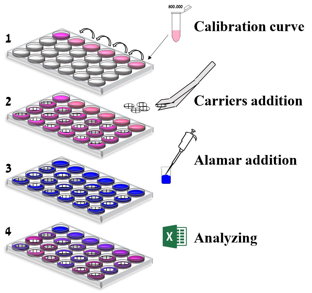
The technology of cell carriers was developed as a response to the need for high cell density to enable higher production levels in cell-based production processes. To follow the production process, quantifying the number of cells on these carriers is required, as well as tracking their viability and proliferation. However, owing to various carriers’ unique structures, tracking the cells is challenging using current traditional assays that were originally developed for monolayers of adherent cells. The current "gold standard" method is counting cell nuclei, which is tedious and counts both live and dead cells. A few other techniques have been developed, but they are all specific to a carrier type and involve specialized equipment. Here, we describe a broad ranging method for counting cells on carriers. The method is based on the Alamar blue dye, a well-known, common marker for cell activity. No separation of the cells from the carriers is needed, nor is any specialized equipment. The method is simple and rapid, and provides comprehensive details necessary for control of production processes in cells. This method can be easily implemented in any cell-based process and other unique platforms for measuring growth of cells.
Graphic abstract:
Schematic of the in situ quantification method.
Background
Cell culture-based production is gaining increasing attention owing to the need for new manufacturing strategies. The advantages of cell-based production processes include the use of defined and serum-free cell cultures that allow greater consistency. Moreover, cell-based processes can be adapted to manufacturing processes involving bioreactors that are scalable and need less space (Aubrit et al., 2015). The mounting use of cell culture-based vaccine production, together with a quest for a more efficient and scalable process, have stimulated the development and implementation of carrier technology. Micro and macrocarriers are small compact surface support matrices for growing adherent cells. Those carriers (from the micrometer to the millimeter size range for micro and macrocarriers, respectively) and their density allows their maintenance in suspension with gentle stirring (Huang et al., 2020).
To better follow and understand a production process, it is necessary to quantify the number of cells on these carriers, as well as to track their viability and proliferation. The gold standard method for quantification of cells on micro and macrocarriers is nuclear staining (Sanford et al., 1951). In this method, cells are separated from the carriers, stained with crystal violet, and counted visually under a microscope. The method is tedious (Berry et al., 1996) and counts both live and dead cells, making it unsuitable for tracking cell condition. As a result, a method that can better quantify cells on micro and macrocarriers is desirable.
Counting cells on carriers poses a challenge to traditional assays due to the carriers’ shape. The main feature of the widely used methods to measure cell quantity (Riss et al., 2013) is dyeing the cells with a fluorescent or colorimetric dye and quantifying the results based on fluorescence or absorbance. Examples of such dyes are glycyl-phenylalanyl-aminofluorocoumarin (GF-AFC), 3-(4,5-dimethylthiazol-2-yl)-2,5-diphenyltetrazolium bromide (MTT), 3-(4,5-dimethylthiazol-2-yl)-5-(3-carboxymethoxyphenyl)-2-(4-sulfophenyl)-2H-tetrazolium (MTS), 2,3-bis-(2-methoxy-4-nitro-5-sulfophenyl)-2H-tetrazolium-5-carboxanilide (XTT), 4-[3-(4-iodophenyl)-2-(4-nitrophenyl)-2H-5-tetrazolio]-1,3-benzene disulfonate (WST-1), and resazurin (Alamar blue). These assays are relatively inexpensive and have a homogeneous format, but their use is limited to single layer cultures (Mosmann, 1983; Berridge and Tan, 1993; Marshall et al., 1995). A limited number of assays for cell quantification on carriers have been developed, but they all require specialized equipment that is not commercially available, and are “matched” with specific micro or macrocarriers (Farrell et al., 2016). Thus, to date, no assays for quantification of cells on carriers that are simple, rapid, reliable, and low-priced are commercially available.
Here, a method for quantification of cells on carriers is described. The assay utilizes Alamar blue, a well-known and common marker for cell activity (Rampersad, 2012), and includes a calibration curve with known cell numbers. When added to cell cultures, the oxidized form of Alamar blue enters the cytosol and is converted to the reduced form by mitochondrial enzyme activity. This redox reaction is accompanied by a shift in the color of the culture medium from indigo blue to fluorescent pink, which can be easily measured by fluorometry (Al-Nasiry et al., 2007). The method can quantify cells in situ on the carriers without requiring separation of cells from the carriers. The method is simple, rapid, elegant, does not require specialized reagents or equipment, and results are strongly correlated with the gold standard assay. The method can be easily adapted for any cell-based process or other unique platforms for cell growth, including additional carriers and other scaffolds for culturing cells such as hydrogels.
Materials and Reagents
1.5 ml Eppendorf tubes (Sarstedt, catalog number: 72.692.005)
Pipette tips (Filtered) (Thermo, catalog numbers: TF112-1000-Q [1,000 µl], T104RS-Q [10 µl], TF140-200-Q [200 µl])
24-well clear plates (Corning, catalog number: 3524-ND)
75 cm2 T-flasks (Grenier, catalog number: 658-975)
500 ml vacuum filter bottle system, Sterile (Corning, catalog number: 431097)
Counting vessels (Invitrogen, catalog number: C10283)
Aluminum foil (Nidix)
Vero cells (WHO, catalog number: RCB 10-87)
Note: Cells used for the calibration curve must be the same as cells on carriers.
L-alanine-L-glutamine (Biological Industries, catalog number: 03-022-1B)
NutriVero FLEX-20 (Biological Industries, catalog number: 05-069-1A)
Pen-strep antibiotics (Biological Industries, catalog number: 03-031-1C)
Phosphate buffered saline (PBS) (Biological Industries, catalog number: 02-020-1A)
Recombinant trypsin-EDTA solution (Biological Industries, catalog number: 03-079-1A)
Trypan blue solution (Biological Industries, catalog number: 03-102-1B)
Alamar blue (Promega, catalog number: G808)
Cell medium (see Recipes)
Equipment
Sterile tweezers (United Scientific Supplies, catalog number: 20A00R146)
Sterile scalpel (Asp medical, catalog number: LDT455)
Pipettors 200 and 1,000 µl (ThermoFisher Scientific, catalog numbers: 4652140, 4652080)
Incubator at 37°C with 5% CO2 (ThermoFisher Scientific, model: Heracell 150i)
Laminar flow hood (ThermoFisher Scientific, model: Msc-advantage)
Refrigerator 4°C (Mondial Elite, model: BEV PV 40 MED)
Centrifuge (Eppendorf, model: Minispin plus spin)
SPARK plate reader (NEOTEC BIO, TECAN)
Cytometer cell countess II (Invitrogen, model: AMQAX1000)
Heater (Talboys, microplate heater)
Software
Microsoft Excel 2010
Procedure
文章信息
版权信息
© 2021 The Authors; exclusive licensee Bio-protocol LLC.
如何引用
Rosen, O., Jayson, A., Natan, N. and Epstein, E. (2021). A Simple Method for in situ Quantification of Cells on Carriers. Bio-protocol 11(23): e4254. DOI: 10.21769/BioProtoc.4254.
分类
细胞生物学 > 基于细胞的分析方法
细胞生物学 > 单细胞分析 > 细胞计数
您对这篇实验方法有问题吗?
在此处发布您的问题,我们将邀请本文作者来回答。同时,我们会将您的问题发布到Bio-protocol Exchange,以便寻求社区成员的帮助。