

Visual-stimuli Four-arm Maze test to Assess Cognition and Vision in Mice
视觉刺激四臂迷宫测试评估小鼠的认知和视觉
发布: 2021年11月20日第11卷第22期 DOI: 10.21769/BioProtoc.4234 浏览次数: 5284
评审: Soyun KimXiaoyu LiuDavid F Delotterie
参见作者原研究论文
The authors used this protocol in:

Jan 2021


Abstract
Visual impairments, notably loss of contrast sensitivity and color vision, were documented in Alzheimer’s disease (AD) patients yet are critically understudied. This protocol describes a novel visual-stimuli four-arm maze (ViS4M; also called visual x-maze), which is a versatile x-shaped maze equipped with spectrum- and intensity-controlled light-emitting diode (LED) sources and dynamic grayscale objects. The ViS4M is designed to allow the assessment of color and contrast vision along with locomotor and cognitive functions in mice. In the color testing mode, the spectral distributions of the LED lights create four homogenous spaces that differ in chromaticity and luminance, corresponding to the mouse visual system. In the contrast sensitivity test, the four grayscale objects are placed in the middle of each arm, contrasting against the black walls and the white floors of the maze. Upon entering the maze, healthy wild-type (WT) mice tend to spontaneously alternate between arms, even under equiluminant conditions of illumination, suggesting that cognitively and visually intact mice use both color and brightness as cues to navigate the maze. Evaluation of the double-transgenic APPSWE/PS1ΔE9 mouse model of AD (AD+ mice) reveals substantial deficits to alternate in both color and contrast modes at an early age, when hippocampal-based memory and learning is still intact. Profiling of timespan, entries, and transition patterns between the different arms uncovers variable aging and AD-associated impairments in color discrimination and contrast sensitivity. The analysis of arm sequences of alternation reveals different pathways of exploration in young WT, old WT, and AD+ mice, which can be used as color and contrast imprints of functionally intact versus impaired mice. Overall, we describe the utility of a novel visual x-maze test to identify behavioral changes in mice related to cognition, as well as color and contrast vision, with high precision and reproducibility.
Graphic abstract:
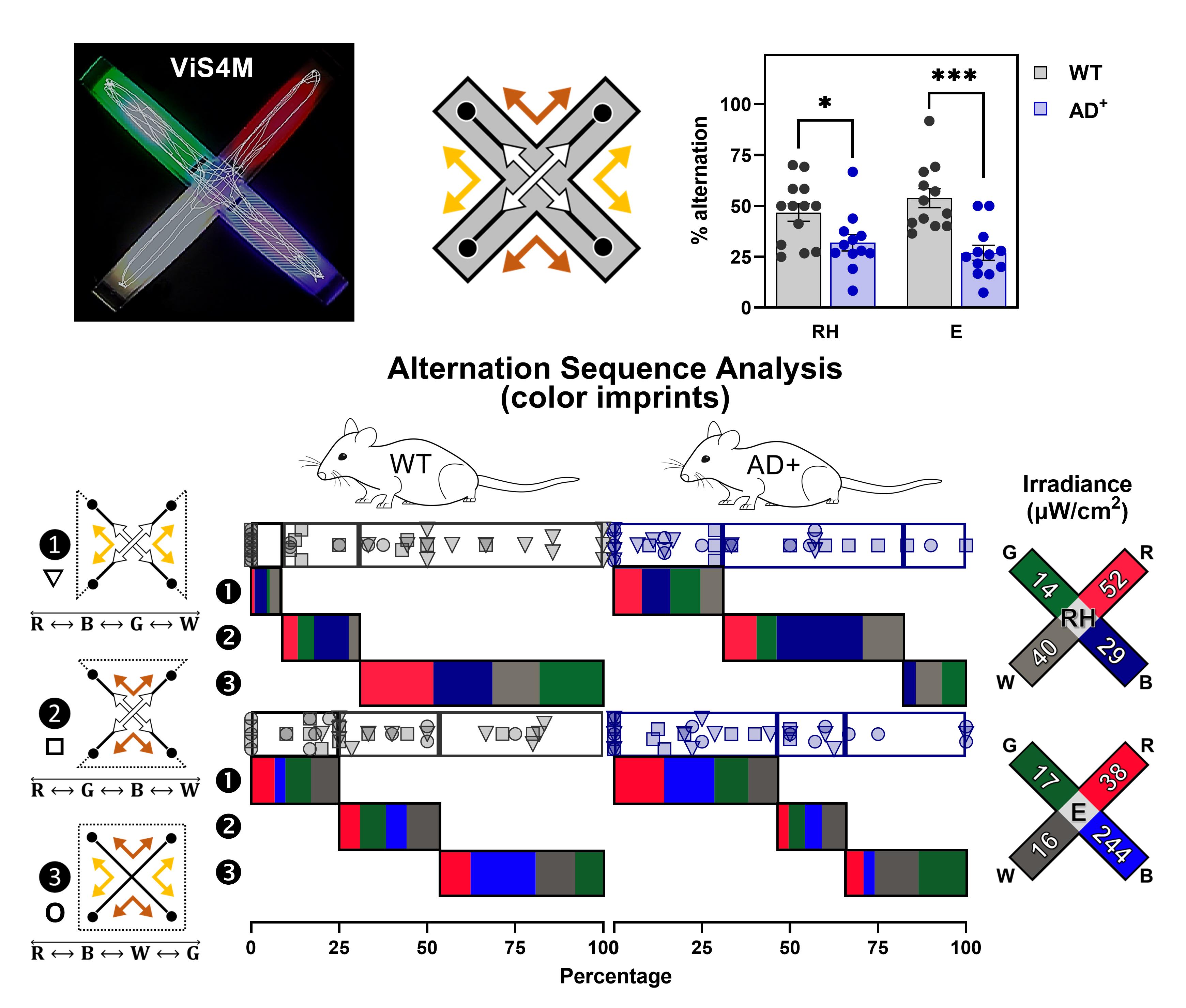
Exploratory behavior of AD+ mice versus age- and sex-matched WT mice is tracked (top left: trajectory from a 5-min video file) in a novel visual-stimuli four-arm maze (ViS4M; also named visual x-maze) equipped with spectrum- and intensity-controlled LED sources or grayscale objects. Consecutive arm entries reveal that APPSWE/PS1ΔE9 (AD+) mice alternate less between arms, as opposed to WT mice. Sequence analysis, according to the three alternation pathways (depicted by white, yellow, and brown arrows) under different conditions of illumination, uncovers specific deficits linked to color vision in AD+ mice, evidenced by a color imprint chart.
Background
Mounting evidence indicates that the pathology of Alzheimer’s Disease (AD) is not restricted to the brain but also extends to the retina. Multiple studies have shown that the retina of mild cognitively impaired (MCI) and AD patients exhibit the neuropathological hallmarks of AD – amyloid β-protein (Aβ) plaques and hyperphosphorylated tau – along with gliosis, vascular dysfunctions, and neuronal degeneration (Blanks et al., 1996a and 1996b; Koronyo-Hamaoui et al., 2011; La Morgia et al., 2016; Koronyo et al., 2017; Dumitrascu et al., 2020; Lee et al., 2020; Lemmens et al., 2020; Shi et al., 2020b; Dumitrascu et al., 2021; Shi et al., 2021; Ngolab et al., 2021; Tadokoro et al., 2021). Moreover, retinal pathology appears to mirror the disease in the brain (Koronyo-Hamaoui et al., 2011; La Morgia et al., 2016; Koronyo et al., 2017; Doustar et al., 2020, Dumitrascu et al., 2020; Shi et al., 2020a and 2020b; Risacher et al., 2020). Clinically, both color and contrast sensitivity were significantly impaired in AD patients when compared with healthy individuals (Chang et al., 2014; Polo et al., 2017). Indeed, visual impairments are among the earliest symptoms documented in these patients, especially loss of contrast sensitivity (Crow et al., 2003; Risacher et al., 2013; Javaid et al., 2016; Salobrar-García et al., 2019) and altered color vision reminiscent of tritanomaly, an abnormality of blue-sensitive retinal cones (Cronin-Golomb et al., 1993; Wijk et al., 1999; Salobrar-García et al., 2019).
Sporadic and transgenic animal models of AD recapitulate AD pathology in the retina and present Aβ deposits and tauopathy that are linked with inflammation, vasculopathy, and neurodegeneration (Perez et al., 2009; Koronyo-Hamaoui et al., 2011; Koronyo et al., 2012; Hart et al., 2016; Doustar et al., 2017; Grimaldi et al., 2018; Hampel et al., 2018; Georgevsky et al., 2019; Chibhabha et al., 2020, Doustar et al., 2020; Habiba et al., 2020; Shi et al., 2020a; Shi et al., 2021). Transgenic mouse models of AD also show disturbances in the visual system, reduced function of ganglion cells and photoreceptors, as well as reduction of inner retinal thickness and atrophy of the visual cortex (Criscuolo et al., 2018; Chiquita et al., 2019). However, the specific visual changes in color and contrast vision have not been previously examined in transgenic mouse models of AD.
Behavioral assessment of color vision and contrast sensitivity in C57BL/6 mice has been previously conducted by optomotor response (Sinex et al., 1979; Prusky et al., 2004), optokinetic reflex (van Alphen et al., 2009), and forced-choice procedures. The latter includes the visual water task (Jacobs et al., 2004; Prusky and Douglas, 2004), psychometric curves in freely moving mice (Busse et al., 2011), and the visual-stimulation environment (Denman et al., 2018). Forced-choice paradigms require days of training and thousands of trials, which are less suitable for aged mice, especially for sensitive, high-attrition rate AD-model mice.
We created the behavioral visual-stimuli four-arm maze (ViS4M or visual x-maze), a novel, controlled, and user-friendly behavioral apparatus specialized for the detection of vision changes in mice (Vit et al., 2021). Unlike previous behavioral tests, the ViS4M is highly sensitive and reproducible, allowing mice to move freely without introducing stress. Importantly, the test relies entirely on innate exploratory behavior and does not require a pre-training phase nor rewards. Moreover, this test can simultaneously assess locomotor, cognitive, and visual functions. We investigated color and contrast sensitivity in the double-transgenic APPSWE/PS1∆E9 (AD+) mice. These AD-model mice and wild-type (WT) mice were tested at three different ages, during six sessions (5 min each) of testing, in five color modes (five conditions of illumination), and one contrast mode. Using the novel ViS4M, we identified early and progressive impairments in color vision and contrast sensitivity in AD-model mice.
In the present manuscript, we provide a detailed description and step-by-step configuration of the ViS4M and accessories, with numerous notes and tips in the equipment and procedure sections. In the data analysis section, we review all the parameters analyzed in the original study (total entries, timespan, alternation, and transitions) with their calculations and supported by relevant examples. We also present an additional analysis of alternation arm sequences and make freely available our own ViS4M Toolbox V1, which includes two Excel spreadsheets to automatically calculate all the aforementioned parameters upon inputting a sequence of arm entries in color and contrast modes. The comprehensive analysis of alternation sequences, as well as unidirectional and bidirectional transitions in combination with linear regressions and visualization tools, allow a better understanding of the locomotor, cognitive, and visual functions of mice measured in the ViS4M.
Materials and Reagents
Animals
Double transgenic B6.Cg-Tg(APPSWE/PSEN1∆E9)85Dbo/Mmjax hemizygous (AD+) mouse strain [RRID:MMRRC_034832-JAX], initially purchased from the Mutant Mouse Resource and Research Center (MMRRC) at the Jackson Laboratory, then bred and maintained at Cedars-Sinai Medical Center.
Non-transgenic wild-type (WT) littermates (Jackson Laboratory, catalog number: 000664)
The mouse colony is housed in a humidity- and temperature-controlled (21-22°C) vivarium on a 12:12-h light/dark cycle (lights on at 8:00 am; lights off at 8:00 pm) with free access to food and water.
Three different cohorts of mice are tested, each representing a different age: 8.5-month-old WT (n=13; 9 males and 4 females) and AD+ (n=12; 9 males and 3 females), 13-month-old WT (n=11; 6 males and 5 females) and AD+ (n=11; 4 males and 7 females), and 18-month-old WT (n=19; 11 males and 8 females) and AD+ (n = 9; 5 males and 4 females).
Note: The double-transgenic APPSWE/PS1ΔE9 is a well-established mouse strain, with early-onset pathology of AD in the hippocampus and cortex (β-amyloid plaques, severe astrogliosis, and microgliosis, as well as brain plasticity deficits and synaptic loss). We and others also reported AD manifestation in the retina of these mice (Ning et al., 2008; Koronyo et al., 2012; Mirzaei et al., 2019; Doustar et al., 2020, Shi et al., 2020a and 2020b). Importantly, cognitive deficits in these mice have been well documented and reproducible. We developed the ViS4M to further characterize visual function, especially color vision and contrast sensitivity. AD pathology in the retina of other mouse strains has been confirmed, such as the aggressive-phenotype 5xFAD or the triple-transgenic 3xTg mice (Criscuolo et al., 2018; Grimaldi et al., 2018; Habiba et al., 2020). This protocol could be implemented on different AD mouse strains, normal aging, and other neurodegenerative models to evaluate visual function.
Equipment
Visual-stimuli four-arm maze (ViS4M) apparatus
Note: The visual-stimuli four-arm maze (ViS4M or Visual X-maze) that we describe below was conceived and developed in-house by our team. The prototype used to collect the present data shares the same specifications (including the color LED lights) with the now commercially available ViS4M manufactured by Maze Engineers (https://conductscience.com/maze/portfolio/visual-x-maze-vis4m/).
Backbone of apparatus
Custom-made x-shaped enclosure built with 15 cm-high black plexiglass walls attached to a glass base
Each arm is perpendicular to the two adjacent arms and is 45 cm long and 10 cm wide (Figure 1A-1B).
Removable transparent floor plates that can be installed at two different levels (6 cm or 11 cm above the glass base) in each arm and in the center of the ViS4M (Figure 1C)
White translucent acrylic plates that can be inserted below each floor level by sliding within small track brackets made of plexiglass (Figure 1C-1D)
Note: Behavioral testing is carried out either on the upper level (color mode) or the lower level (contrast mode) but not simultaneously on both levels.
The center of the apparatus is a neutral area with no white translucent plate and no light source directly below it (Figure 1A-1C)
Plastic transparent covers with perforated holes to help with breathing
Note: Color vision and contrast sensitivity are different aspects of vision that involve different photoreceptors. While the contrast mode involves the function of rods (lower mesopic range), the color mode requires functional cones (transition between mesopic and photopic range). These two modalities of testing are complementary to appreciate the functionality of rod and cone photoreceptors.
Light-emitting diode (LED) lights (for color mode)
Each LED strip source consists of an array of surface-mounted device (SMD) 3528 LED chips evenly spaced in four rows (27 LED chips per row) and is individually inserted in each arm of the ViS4M, directly onto the glass base (Figure 1D). Spectra of the light sources were determined using an Ocean Optics USB2000 spectrometer and further validated with a Sekonic C700-U spectrometer (Figure 1E).
Red monochromatic light: wavelength (λ) = 628 nm, full width at half maximum (FWHM) = 17 nm
Green monochromatic light: λ = 517 nm, FWHM = 31 nm
Blue monochromatic light: λ = 452 nm, FWHM = 22 nm
White light: λ1 = 441 nm, FWHM = 19 nm and λ2 = 533 nm, FWHM = 104 nm
The white LED is made of a blue-emitting diode that also excites a yellow-emitting phosphor [cerium doped yttrium aluminum garnet (Ce:YAG-Y3Al5O12) crystals] embedded in the epoxy dome.
Note: The criterion for choosing the LED colors is as follows: the red light as a dark-space control arm with low- to no-color stimulus; the green light to stimulate the mouse retinal M-opsin in M-cones, without stimulating S-opsin; and the blue light to stimulate the mouse retinal S-opsin in S- and M-cones, in addition to the M-opsin.
The following components control the brightness of the light stimuli.
LED single color dimmers using pulse width modulation (PWM) technology
Individual remote devices to control the dimmers
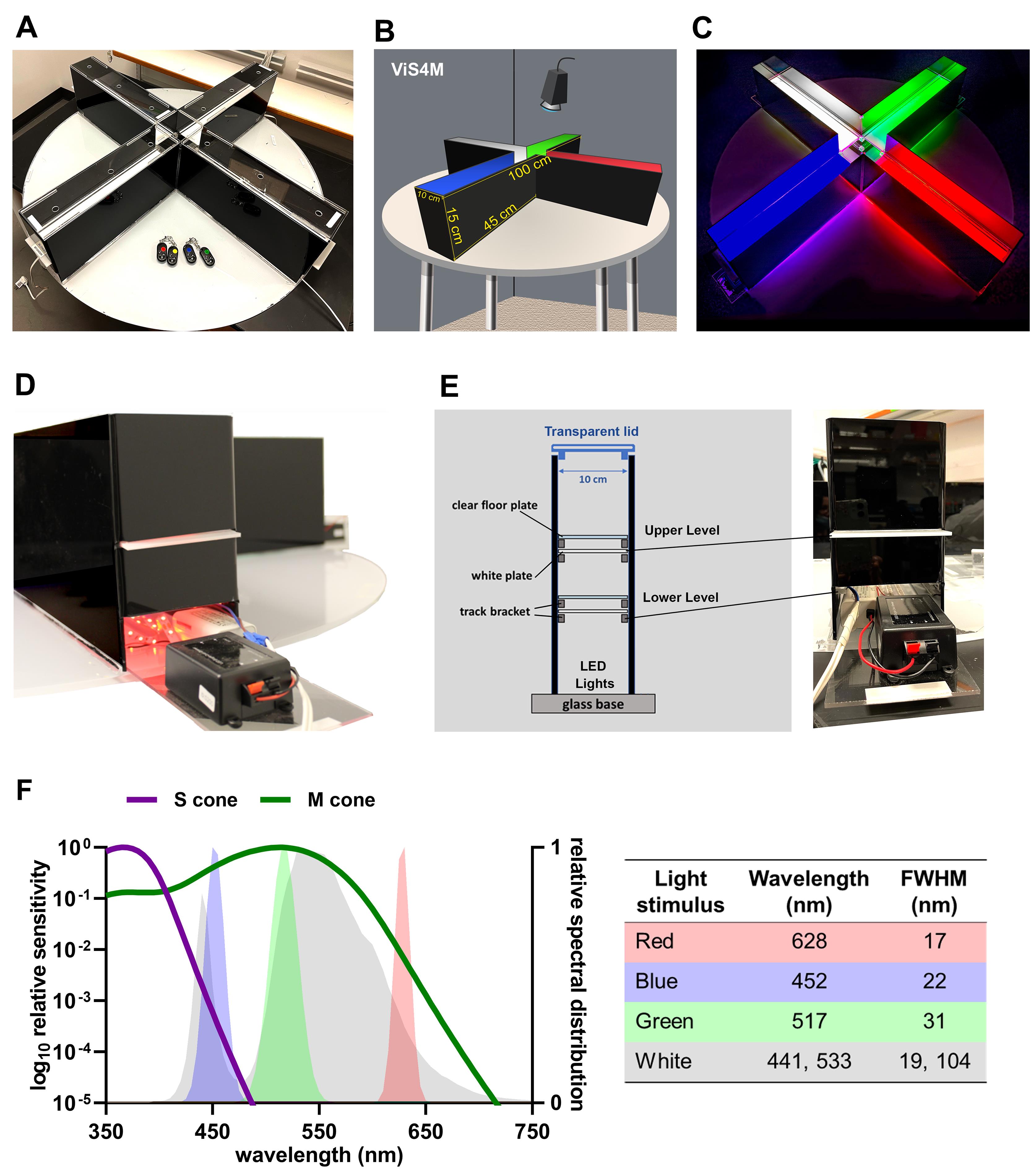
Figure 1. Characteristics of the ViS4M in color mode. (A) Photograph and (B) illustration of the x-shaped ViS4M in color mode with apparatus measurements. (C) Photograph of the ViS4M in color mode, E condition, showing the illuminated arms. (D) Photograph of an arm with the opening for the red lights and with the white translucent plate inserted at the upper level. (E) Illustration of the positioning of the clear floor and white translucent diffuser plates within the arms of the maze, paired with a photograph of the outside front-view of an arm. (F) Spectral distribution of the LED sources according to mouse M- and S-opsins sensitivity with table showing the characteristics of the light stimuli (wavelength, FWHM).Grayscale objects (for contrast mode)
Four objects have the following characteristics (Figure 2A).
Identical shape of a right-angled triangular prism
Dimensions: base (adjacent side) = 5 cm; height (opposite side) = 1 cm
Different shades (black, gray, white, and clear) that create different contrasts with the black walls and white floors of the maze (Figure 2B)
The luminance ratios of the objects against the black walls and/or white floors as measured with a light meter are indicated below.
Black object against the black walls = 1.06 (minimal to no contrast)
Gray object against the black walls = 6.00
White object against the black walls = 9.69 (high contrast against the black walls but minimal contrast with the white floor)
Clear object against the black walls = 6.56
Flexible positioning and location for the placement of the grayscale objects to accommodate investigator’s goals
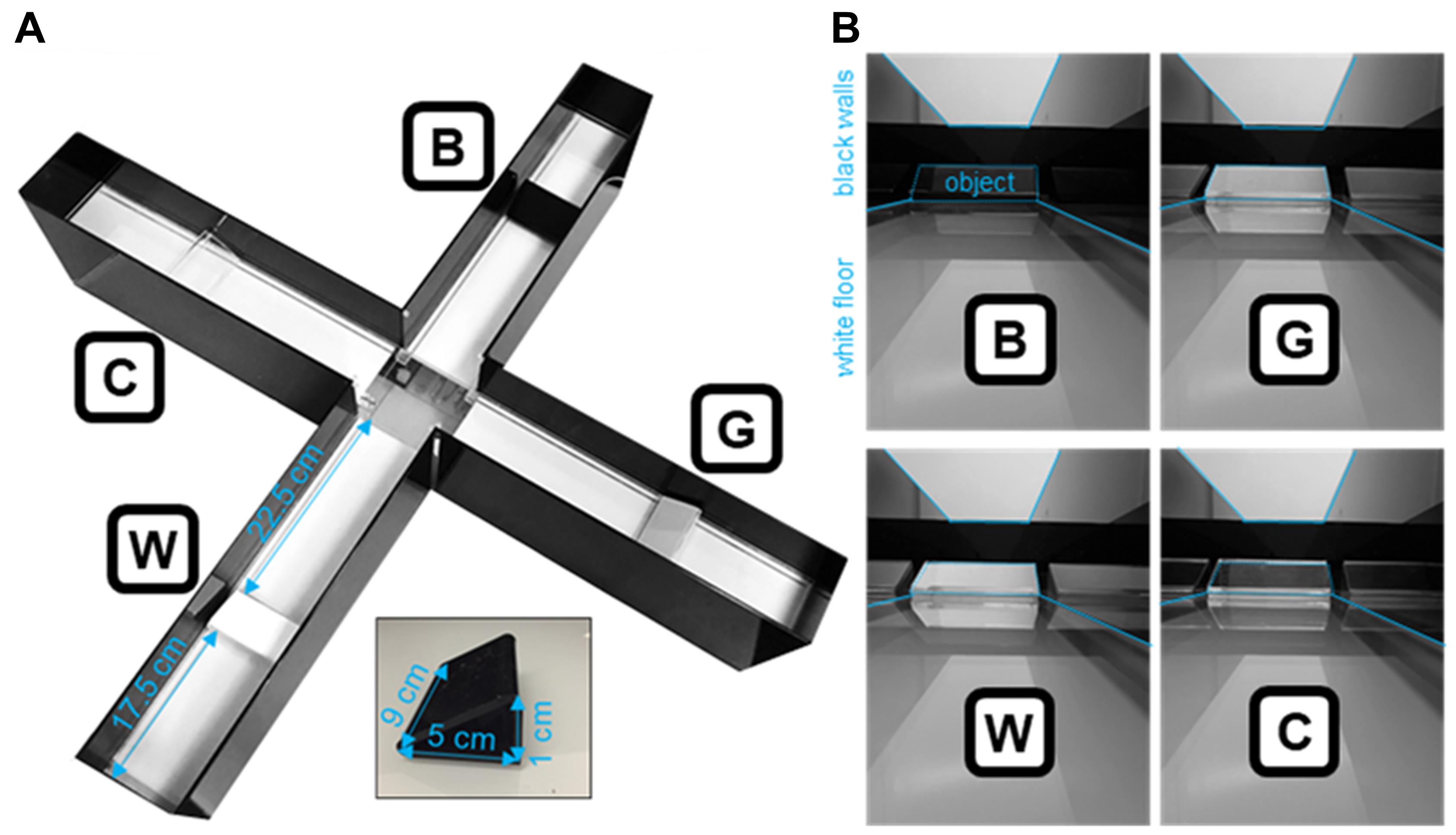
Figure 2. Characteristics of the ViS4M in contrast mode. (A) Photograph of the ViS4M in contrast mode with measurements for positioning of the grayscale objects. (B) Photographs of the objects within each arm as seen from the center of the ViS4M. Objects, black walls, and white floors are delineated to better assess contrasts. (A-B) B, Black; C, Clear; G, Gray; W, White.
Other accessories
Sekonic Flashmate L-308S light meter (Sekonic, catalog number: 401-309)
Timer
Digital USB camera
Note: A color camera is preferable to identify the colored arms. However, a black and white camera can be used, provided the position of each arm on the screen is recorded. A short clip of the apparatus positioning can be made prior to each testing session with the lights of the room turned on and the arms identified by name.
USB camera varifocal lens with 2.8-12 mm focal length
Software
Rodent Toolbox V1 (freely available at https://www.ndcn.ox.ac.uk/team/stuart-peirson)
ANY-maze behavior tracking software 6.3 (www.stoeltingco.com/anymaze/video-tracking/software.html) or other video tracking software, such as EthoVision XT (www.noldus.com/ethovision-xt
ViS4M Toolbox V1 (Supplementary file: ViS4M Toolbox V1)
GraphPad Prism 9.0
ParallelSets V2 (freely available at Parallel Sets (eagereyes.org))
Circos online (freely available at mkweb.bcgsc.ca/tableviewer/)
Procedure
文章信息
版权信息
© 2021 The Authors; exclusive licensee Bio-protocol LLC.
如何引用
Vit, J., Fuchs, D., Angel, A., Levy, A., Lamensdorf, I., Black, K. L., Koronyo, Y. and Koronyo-Hamaoui, M. (2021). Visual-stimuli Four-arm Maze test to Assess Cognition and Vision in Mice. Bio-protocol 11(22): e4234. DOI: 10.21769/BioProtoc.4234.
分类
神经科学 > 感觉和运动系统 > 视觉系统
神经科学 > 神经系统疾病 > 动物模型
您对这篇实验方法有问题吗?
在此处发布您的问题,我们将邀请本文作者来回答。同时,我们会将您的问题发布到Bio-protocol Exchange,以便寻求社区成员的帮助。