

Whole-mount Immunohistochemistry to Visualize Mouse Embryonic Dermal Lymphatic Vasculature
小鼠胚胎真皮淋巴血管的免疫组织化学观察
发布: 2021年10月20日第11卷第20期 DOI: 10.21769/BioProtoc.4186 浏览次数: 2694
评审: Giusy TornilloFrancesca Mingozzi
参见作者原研究论文
The authors used this protocol in:
Dec 2020
Advertisement


Abstract
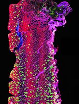
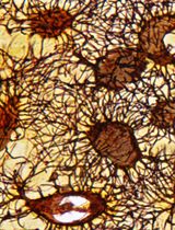
Lymphatic vessels are abundant in the skin where they regulate interstitial fluid uptake and immune surveillance. Defects in dermal lymphatic vessels, such as fewer vessels and abnormal lymphatic vessel coverage with mural cells, are frequently associated with lymphedema and other lymphatic disorders. Whole-mount immunohistochemistry allows the visualization of dermal lymphatic vessels and identifies morphogenetic defects. Most dermal lymphatic vessels start growing during embryogenesis from lymph sacs that are located close to the axilla towards the dorsal and ventral midlines. Here, we present an approach that we have developed to permeabilize, immunolabel, clear, and visualize the lymphatic vessels. These simple and inexpensive techniques reproducibly generate images of dermal lymphatic vessels with great clarity.
Keywords: Lymphatic vessels (淋巴管)Background
Lymphatic vessels are 3-dimensional structures that regulate interstitial fluid homeostasis, lipid absorption, and immune cell trafficking. Defects in lymphatic vessels are associated with diseases such as lymphedema and lymphatic malformations and could result in tissue swelling and inflammation. The growth and patterning of dermal lymphatic vessels can be visualized by whole-mount immunohistochemistry. This approach also identifies defects in lymphatic vessels such as fewer branch points, lack of lymphatic valves, and abnormal recruitment of mural cells. Nevertheless, whole-mount immunohistochemistry is a challenging procedure as skin is a cell-dense tissue with plenty of fat and extracellular matrix proteins such as collagen. To overcome this limitation, we have combined the existing approaches to harvest and visualize dermal lymphatic vessels with the immunolabeling-enabled three-dimensional imaging of solvent-cleared organs (iDISCO) protocol to obtain superior antibody permeabilization and tissue clearing (James et al., 2013; Renier et al., 2014; Betterman et al., 2018). This inexpensive approach is simple and easy to perform. High quality images can be obtained within a week of starting the procedure. Here, we present the step-by-step protocol to analyze the dermal lymphatic vessels of mouse embryos (embryonic day (E) 14.5-18.5). However, this protocol can be used with minimal modifications to visualize blood and lymphatic vessels in other organs, such as the embryonic and postnatal mesentery and adult ear (Cha et al., 2016, 2018 and 2020; Geng et al., 2016 and 2020; Mahamud et al., 2019).
Materials and Reagents
12-well plate
6-well plate
24-well plate
Sylgard 184 silicone elastomer kit (Fisher Scientific, catalog number: NC9285739)
Insect pin (Fine Science Tools, catalog number: 26002-20)
Heparin (Millipore Sigma, catalog number: H3393): resuspend in water at 50 mg/ml, aliquot, and store at -20°C)
Sodium deoxycholate (VWR, catalog number: 0613-100g): to make 10% stock solution dissolve 5 g in 30 ml water, add water to 50 ml, and store at room temperature protected from light.
Microscope slides (Denville Scientific INC, catalog number: M1021)
Wax pen (Vector, catalog number: H-4000)
Microscope cover glass (Fisher Scientific, catalog number: 12-545-M)
Mounting medium (Electron Microscopy Sciences, catalog number: 17985-01-02-03)
Goat anti mVEGFR3 (R&D System, catalog number: AF743)
Rat anti mCD31 (BD Pharmingen, catalog number: 553370)
Rabbit anti PROX1 (AngioBio Co. catalog number: 11-002)
Rat anti mNRP2 (R&D System, catalog number: AF567)
Goat anti hPROX1 (R&D System, catalog number: AF2727)
Goat anti mVEGFR2 (R&D System, catalog number: AF644)
Goat anti mLYVE-1 (R&D System, catalog number: AF2125)
Sheep anti mFOXC2 (R&D System, catalog number: AF6989)
Rabbit anti mClaudin 5 (ThernoFisher, catalog number: 34-1600)
CY3-conjugated Mouse anti-Rabbit (Jackson ImmunoResearch Laboratories, catalog number: 711-165-152)
Alexa488-conjugated Donkey anti-Goat (Jackson ImmunoResearch Laboratories, catalog number: 705-545-147)
CY5-conjugated Donkey anti-Rat (Jackson ImmunoResearch Laboratories, catalog number: 712-175-150)
CY3-conjugated Donkey anti-Sheep (Jackson ImmunoResearch Laboratories, catalog number: 713-165-147)
Rat anti mCD144 (BD Pharmingen, catalog number: 550548)
20× PBS (see Recipes)
20% paraformaldehyde (PFA) (see Recipes)
10% Triton X-100 (see Recipes)
PBST (see Recipes)
PBST/20% DMSO (see Recipes)
10% Tween 20 (see Recipes)
10% NP40 (see Recipes)
PTTDND (see Recipes)
1 M glycine (see Recipes)
PTDG (see Recipes)
Blocking buffer (see Recipes)
PTWH (see Recipes)
PTWH/5% DMSO/3% donkey serum (see Recipes)
PTWH/3% donkey serum (see Recipes)
Equipment
Forceps (Fine Science Tools, catalog number: 11252-00)
Benchtop rocker
Spring scissors (Fine Science Tools, catalog number: 15000-08))
Nikon microscope
Zeiss confocal microscope
Procedure
文章信息
版权信息
© 2021 The Authors; exclusive licensee Bio-protocol LLC.
如何引用
Geng, X. and Srinivasan, R. S. (2021). Whole-mount Immunohistochemistry to Visualize Mouse Embryonic Dermal Lymphatic Vasculature. Bio-protocol 11(20): e4186. DOI: 10.21769/BioProtoc.4186.
分类
发育生物学 > 形态建成 > 器官形成
细胞生物学 > 组织分析 > 组织成像
生物科学 > 生物技术
您对这篇实验方法有问题吗?
在此处发布您的问题,我们将邀请本文作者来回答。同时,我们会将您的问题发布到Bio-protocol Exchange,以便寻求社区成员的帮助。