

Microscopic Detection of ASC Inflammasomes in Bone Marrow Derived Macrophages Post Stimulation
刺激后骨髓源性巨噬细胞中ASC炎症小体的显微检测
发布: 2021年09月05日第11卷第17期 DOI: 10.21769/BioProtoc.4151 浏览次数: 4210
评审: Andrea PuharVishal NehruAnonymous reviewer(s)
参见作者原研究论文
The authors used this protocol in:

Dec 2020


Abstract
An inflammasome is an intracellular multiprotein complex that plays important roles in host defense and inflammatory responses. Inflammasomes are typically composed of the adaptor protein apoptosis-associated speck-like protein containing a CARD (ASC), cytoplasmic sensor protein, and the effector protein pro-caspase-1. ASC assembly into a protein complex termed ASC speck is a readout for inflammasome activation. Here, we provide a step-by-step protocol for the detection of ASC speck by confocal microscopy in Bone marrow derived macrophages (BMBDs) triggered by chemical stimuli and bacterial pathogens. We also describe the detailed procedure for the generation of BMDMs, stimulating conditions for inflammasome activation, immunofluorescence cell staining of ASC protein, and microscopic examination. Thus far, this method is a simple and reliable manner to visualize and quantify the intracellular localization of ASC speck.
Graphic abstract:
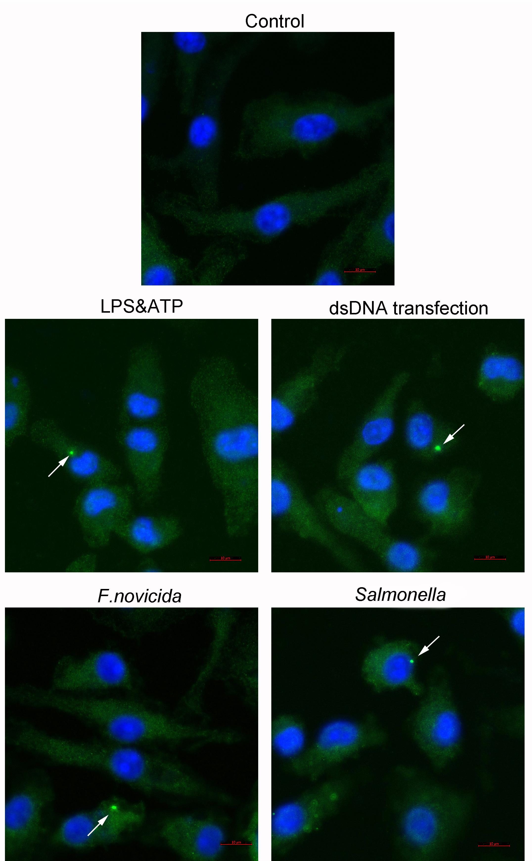
Figure 1. Confocal microscopy detection of ASC speck formation in untreated WT BMDMs and WT BMDMs stimulated with LPS and ATP, transfected with dsDNA, and infected with F. novicida or Salmonella as indicated. Arrow indicates the ASC speck. Scale bars: 10 μm.
Background
The innate immune system has a key role in initiating and orchestrating host defense by detecting invading pathogens through membrane-bound and cytosolic pattern recognition receptors (PRRs), which recognize pathogen-associated and damage-associated molecular patterns (PAMPs and DAMPs). Inflammasome activation is an essential innate immune event in response to pathogenic infection and sterile stimuli that causes the initiation of pyroptotic cell death and the release of the proinflammatory cytokines IL-1β and IL-18. Inflammasome assembly is triggered by the activation of upstream sensors, such as NLRP1, NLRP3, AIM2, NLRC4, and PYRIN. Upon activation, sensor proteins form a complex in an ASC-dependent and -independent manner to mediate caspase-1 cleavage and activation. Cleaved caspase-1 in turn leads to the maturation of proinflammatory cytokines IL-1β and IL-18 and the process of GSDMD-mediated pyroptosis (Rathinam and Fitzgerald, 2016). The adaptor ASC protein is composed of a PYRIN domain (PYD) and caspase recruitment domain (CARD), which help ASC function as an adaptor to interact with upstream sensor and effector caspase-1 (Agrawal and Jha, 2020). ASC speck formation is a hallmark of inflammasome activation. Confocal microscopy and flow cytometry are two major methods to detect ASC speck formation (Stutz et al., 2013; Sester et al., 2015; Beilharz et al., 2016; Hoss et al., 2018).
We performed and published the determination of ASC speck formation in BMDMs after the activation of NLRP3, AIM2, and NLRC4 inflammasomes (Guo et al., 2020). In comparison with the method using ASC-GFP fusion protein and flow cytometry, this procedure is able to detect endogenous ASC speck formation and can visualize and quantify subcellular localization of the inflammasome complex with the help of cellular organelle staining. This protocol can be utilized to evaluate any ASC speck formation in other cells after ASC-dependent inflammasome activation.
Materials and Reagents
Microscope Slides (Citotest, catalog number: 198105)
12-well plate (Jet, catalog number: TCP011012)
10 cm cell culture dish (Jet, catalog number: TCD010100)
15/50 ml sterile centrifuge tube (Jet, catalog number: CFT011500)
Serological Pipet (JETBIOFIL, catalog number: GSP-010-005/GSP-010-010)
25 cm Cell Scraper (BIOFIL, catalog number: CSC011025)
Microscope Cover Glass (NEST, catalog number: 801008, Φ15 mm)
5 ml syringe with 26 G needle
20 ml syringe with 18 G needle
Mice (C57/BLJ6, Beijing Vital River Laboratory Animal Technology Co., Ltd)
RPMI 1640 (Gibco, catalog number: 31800022, 4°C)
DMEM/F12 (Gibco, catalog number: 12500062, 4°C)
FBS (Hyclone, catalog number: SH30084.03, -20°C/4°C)
Nonessential amino acids (Gibco, catalog number: 111140-050, 4°C)
Penicillin-streptomycin (Gibco, catalog number: 15140-122, 4°C)
PBS (Gibco, catalog number: 21600-069, 4°C)
4% paraformaldehyde (PFA) (Sangon Biotech, catalog number: E672002-0500, 15-25°C (RT))
BSA (Sangon Biotech, catalog number: A500023, 4°C)
Anti-ASC (AdipoGen, catalog number: AG-25B-0006, -20°C)
Saponin (Sigma, catalog number: 47036, 4°C)
Fluorescence conjugated secondary antibody (Alexa FluorTM 488 goat anti-Rabbit) (Invivogen, catalog number: A11008, -4°C)
LPS (Invivogen, catalog number: tlrl-smlps, -20°C)
ATP (Sigma, catalog number: FLAAS, -20°C)
X-fect Transfection Reagent (X-fect polymer, X-fect buffer) (Clontech, catalog number: 631318, -20°C)
4’,6-diamidine-2-phenylindole dihydrochloride (DAPI) (CST, catalog number: 8961 S, -20°C)
Anti-fluorescence attenuation sealant (Solarbio, catalog number: S2100, 4°C)
TWEEN® 20 (Sigma-Aldrich, catalog number: P2287)
Cell-neubauer improved (LW Scientific, 0.0025 mm2)
BBLTM TrypticaseTM Soy Broth (BD, catalog number: 211768, RT)
NaCl (Sangon Biotech, catalog number: A501218-0001, RT)
Tryptone (OXOID, catalog number: LP0042, RT)
Yeast extract (OXOID, catalog number: LP0021, RT)
Agar (SbaseBio, catalog number: A010-1.1, RT)
TSB solid media (see Recipes)
TSB liquid media (see Recipes)
LB solid media (see Recipes)
LB liquid media (see Recipes)
1% BSA (see Recipes)
0.1% saponin (see Recipes)
0.1% PBST (see Recipes)
Equipment
Opthalmic scissors (Beijing Bao Yuan Industrial Technology, catalog number: M-Y003)
Opthalmic forceps (Beijing Bao Yuan Industrial Technology, catalog number: M-Y005)
Confocal Microscope (ZEISS, model: LSM880)
Microscope (ZEISS, model: Primo vert iLED)
Biological Safety Cabinets Clean Benches (ThermoFisher Scientific, model: 1300)
CO2 Incubator (ThermoFisher Scientific, model: 3111)
Portable Pipet-Aid (Drummond, catalog number: 4-000-201)
Diaphragm vacuum pump (Tianjin Jinteng Experiment Equipment, model: GM-0.33A)
Nanophotometer p-class (IMPLEN, model: NT-80)
-80°C freezer (Thermo, model: FDE30086FV)
Software
ZEN black_2-3SP1 (ZEISS, https://www.zeiss.com/corporate/us/home.html)
ZEN blue 2.6 (ZEISS, https://www.zeiss.com/corporate/us/home.html)
Procedure
文章信息
版权信息
© 2021 The Authors; exclusive licensee Bio-protocol LLC.
如何引用
Li, L., Mao, R. and Qi, X. (2021). Microscopic Detection of ASC Inflammasomes in Bone Marrow Derived Macrophages Post Stimulation. Bio-protocol 11(17): e4151. DOI: 10.21769/BioProtoc.4151.
分类
免疫学 > 免疫细胞功能 > 巨噬细胞
微生物学 > 微生物-宿主相互作用 > 细菌
细胞生物学 > 细胞成像 > 共聚焦显微镜
您对这篇实验方法有问题吗?
在此处发布您的问题,我们将邀请本文作者来回答。同时,我们会将您的问题发布到Bio-protocol Exchange,以便寻求社区成员的帮助。