

Oil Red O Staining for Lipid Content in Caenorhabditis elegans
油红O染色测定秀丽隐杆线虫脂质含量
发布: 2021年08月20日第11卷第16期 DOI: 10.21769/BioProtoc.4124 浏览次数: 7751
评审: Demosthenis ChronisMichael EnosAnonymous reviewer(s)
参见作者原研究论文
The authors used this protocol in:

Nov 2020
Advertisement


Abstract
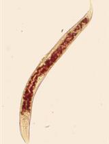
The nematode Caenorhabditis elegans has emerged as a popular model system for studying the regulation of lipid metabolism. Therefore, it is critical to develop a method for determining fat storage in individual worms. Oil Red O (ORO) staining has been validated as an accurate assessment for major fat storage in C. elegans. Here, we describe an optimized protocol for ORO staining of C. elegans and provide detailed instructions for quantifying the intensity of ORO signal in images acquired by light microscopy.
Keywords: Oil Red O (油红O(苏丹红))Background
Recently, there has been a growing interest in studying the interplay between lipid metabolism and energy homeostasis. Caenorhabditis elegans has proved to be a powerful genetic system for elucidating complex physiological mechanisms and identifying novel genetic pathways. Lipid metabolism is a highly conserved process across eukaryotes. Many of the mammalian genes of the lipid metabolic pathways function similarly in C. elegans (Ashrafi, 2007; Jones and Ashrafi, 2009). Furthermore, several lipid regulatory pathways identified in C. elegans have been confirmed in mammalian systems (McKay et al., 2003; Suh et al., 2007). These features have made C. elegans an excellent animal model to study the regulation of fat accumulation and distribution.
To understand the molecular mechanisms regulating fat metabolism, a precise assessment of fat accumulation levels in worms is required. Various methods have been applied to visualize and measure fat content in C. elegans (Yen et al., 2010; Barros et al., 2012). Among them, Oil Red O (ORO) staining of fixed worms has been proved to be an accurate method to assess fat storage in C. elegans (O'Rourke et al., 2009; Zhang et al., 2010). ORO is a fat-soluble diazol dye that detects neutral lipids and cholesteryl esters but not biological membranes (Ramirez-Zacarias et al., 1992). Here, we demonstrate an ORO staining protocol with paraformaldehyde fixation. The method is readily applicable as it only requires standard laboratory and computer equipment. As an example of the application, we have included data on visualization and quantification of fat contents in wild-type N2, daf-2(e1370) mutants, and eat-2(ad1116) mutants.
Materials and Reagents
60 mm Petri dish (Greiner Bio-One, catalog number: 628102)
1.5 ml microcentrifuge tube (AXYGEN, catalog number: MCT-150-C)
Glass slide and coverslip (MARIENFELD, catalog number: 0107052)
10 ml syringe (Terumo, catalog number: DVR-3424)
0.22 μm syringe filters (MCE membrane filter, JET BIOFIL, catalog number: AGC-F-MCE022-13)
C. elegans strains were obtained from Caenorhabditis Genetics Center:
N2 Bristol, eat-2(ad1116), daf-2(e1370)
OP50-1 Escherichia coli bacteria (Caenorhabditis Genetics Center)
Oil Red O stain (Sigma, catalog number: O0625)
Sodium hypochlorite solution (6-14% active chlorine basis; Sigma, catalog number: 13440)
K2HPO4 (J.T. Baker, catalog number: 3252-01)
Bacto agar (BD, catalog number: 214510)
Bacteriological peptone (BD, catalog number: 211677)
CaCl2·2H2O (Sigma, catalog number: C3306)
Cholesterol (Sigma, catalog number: C3045)
EGTA (Sigma, catalog number: E8145)
Spermidine (Sigma, catalog number: S2626)
Spermine (Sigma, catalog number: S4264)
PIPES (Sigma, catalog number: P6757)
2-mercaptoethanol (Sigma, catalog number: M3148)
16% paraformaldehyde (Alfa Asear, catalog number: 43368)
KCl (J.T. Baker, catalog number: 3040-01)
2-Propanol (Sigma, catalog number: I9516)
KH2PO4 (Sigma, catalog number: P9791)
Na2HPO4·7H2O (OMNIPUR, catalog number: 8210)
NaCl (J.T. Baker, catalog number: 3624-05)
MgSO4·7H2O (Sigma, catalog number: M2773)
Triton X-100 (Sigma, catalog number: T8787)
LB broth (BIO-DOC, catalog number: BDLBB-500)
Nematode growth media (NGM) (see Recipes)
2× MRWB stock solution (see Recipes)
M9 buffer (see Recipes)
Nematode growth medium (NGM) agar for 1 L medium (see Recipes)
LB media (see Recipes)
Bleaching solution (see Recipes)
Oil Red O staining stock solution (see Recipes)
Potassium phosphate buffer (see Recipes)
Equipment
Low temperature refrigerated Incubator (Precision, catalog number: PR505755R, temperatures ranging from -10°C to +60°C)
Microcentrifuge (Thermo Scientific, model: Pico 17, 24 × 1.5/2 ml rotor, max. speed 13,300 rpm)
Dissecting microscope (Olympus, model: SZ61)
Fluorescence microscopy (Olympus, model: BX63)
Software
MatLab (The MathWorks, https://www.mathworks.com/products/matlab.html)
Cellsens (Olympus, https://www.olympus-lifescience.com/en/software/cellsens/)
Procedure
文章信息
版权信息
© 2021 The Authors; exclusive licensee Bio-protocol LLC.
如何引用
Wang, F. and Ching, T. (2021). Oil Red O Staining for Lipid Content in Caenorhabditis elegans. Bio-protocol 11(16): e4124. DOI: 10.21769/BioProtoc.4124.
分类
生物化学 > 脂质 > 脂质测定
您对这篇实验方法有问题吗?
在此处发布您的问题,我们将邀请本文作者来回答。同时,我们会将您的问题发布到Bio-protocol Exchange,以便寻求社区成员的帮助。