

Mouse Periosteal Cell Culture, in vitro Differentiation, and in vivo Transplantation in Tibial Fractures
小鼠骨膜细胞培养、体外分化及胫骨骨折的体内移植
(*contributed equally to this work) 发布: 2021年08月05日第11卷第15期 DOI: 10.21769/BioProtoc.4107 浏览次数: 4654
评审: Giusy TornilloJosé M. DiasTakashi NishinaZheng Xu
参见作者原研究论文
The authors used this protocol in:
Oct 2020
Advertisement


Abstract
The periosteum covering the outer surface of bone contains skeletal stem/progenitor cells that can efficiently form cartilage and bone during bone repair. Several methods have been described to isolate periosteal cells based on bone scraping and/or enzymatic digestion. Here, we describe an explant culture method to isolate periosteum-derived stem/progenitor cells for subsequent in vitro and in vivo analyses. Periosteal cells (PCs) isolated using this protocol express mesenchymal markers, can be expanded in vitro, and exhibit high regenerative potential after in vivo transplantation at a fracture site, suggesting that this protocol can be employed for PC production to use in new cell-based therapies.
Keywords: Periosteum (骨膜)Background
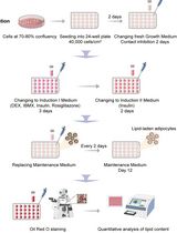
Bone regeneration is a highly efficient process. After bone fracture, skeletal stem/progenitor cells are activated and differentiate into chondrocytes and osteoblasts that form cartilage and bone to consolidate the fracture. Skeletal stem/progenitor cells originate from the bone itself including bone-marrow and periosteum, as well as from adjacent soft tissue. The diverse origins of skeletal stem/progenitor cells during bone regeneration suggest that these cells can be obtained from various sources for stem cell therapies. Due to their accessibility, bone marrow stromal cells (BMSCs) are the most studied (Arthur and Gronthos, 2020); however, their variable osteogenic potential highlights the need for new sources of cells capable of contributing efficiently to the repair process. Recent studies have revealed the role of the periosteum as an essential source of stem/progenitor cells during bone regeneration (Debnath et al., 2018; Duchamp de Lageneste et al., 2018; Ortinau et al., 2019). When transplanted to a bone fracture site, periosteal cells (PCs) display a higher regenerative potential than BMSCs and have the ability to correct a bone repair failure (Duchamp de Lageneste et al., 2018; Julien et al., 2020). Isolating mouse PCs is challenging since the periosteum is a very thin layer of tissue on the outer surface of bone. Several methods have been previously described to isolate PCs in mice, relying on enzymatic digestion and periosteum scraping or peeling (Brownlow et al., 2000; Arnsdorf et al., 2009; Wang et al., 2010; Chang and Knothe Tate, 2012; van Gastel et al., 2012). Here, we describe a method to isolate PCs based on cell migration from bone explants without digestion or periosteum separation from the bone (Figure 1). We developed this method to analyze PC properties for direct comparison with BMSCs that are usually isolated by direct bone marrow flushing and plating. Long bones free of skeletal muscle, epiphyses, and bone-marrow are placed in culture to allow PC migration and proliferation. PCs isolated using this protocol express mesenchymal markers (Figure 2), display in vitro adipogenic, osteogenic, and chondrogenic differentiation capacities (Figure 3), and are able to form cartilage and bone upon in vivo transplantation at the site of a tibial fracture (Figure 4). PCs therefore maintain their osteochondrogenic capacities, offering new potential perspectives for the study of PCs and their use in cell-based therapies.
Materials and Reagents
Falcon® 5-ml round-bottomed polystyrene test tubes (Corning, catalog number: 352235)
40-μm cell strainer (Fisher Scientific, catalog number: 352340)
Conical tubes, 15-ml and 50-ml (Falcon, catalog numbers: 352097 [15 ml] and 352070 [50 ml] or equivalent)
25 G needles (Terumo, catalog number: AN*2516R1)
1-ml syringes (Terumo, catalog number: SS+01H1)
60-mm TPP culture dishes (TPP, catalog number: 93060)
100-mm TPP culture dishes (TPP, catalog number: 93100)
6-well plates (TPP, catalog number: 009206)
Greiner Bio-One Petri dishes (bacterial dish; Dutscher, catalog number: 633185)
10-ml and 25-ml pipets (Dutscher, catalog numbers: 357551 [10 ml] and 357535 [25 ml] or equivalent)
Cell scrapers (TPP, catalog number: 99010 or equivalent)
Kova® slides (Fisher Scientific, catalog number: 22-270141)
Falcon® 5-ml round-bottomed polystyrene test tubes (Corning, catalog number: 352235)
Glass slides, Superfrost Plus (Thermo Fisher, catalog number: J1800AMNZ)
Coverslips (Labellians, catalog number: LCO2460M)
For periosteum-derived cell culture: 4 to 8-week-old mice in the C57BL/6J background (see Note 1)
Hosts for in vivo cell transplantation: 10 to 14-week-old mice in the C57BL/6J background (see Note 2)
DMEM (Life Technologies, catalog number: 11966-025)
Penicillin-streptomycin (P/S; Life Technologies, catalog number:15140-122)
α-Modified Eagle’s Medium (α-MEM with GlutaMAX; Life Technologies, catalog number: 32561-029)
Lot-selected fetal bovine serum (FBS; Life Technologies, catalog number:10270-106)
Recombinant mouse basic fibroblast growth factor (bFGF; R&D, catalog number: 3139FB/CF)
Trypan Blue stain (Life Technologies, catalog number: 15250-061)
Trypsin-EDTA (0.25%), phenol red (Life Technologies, catalog number: 25200056)
Dexamethasone (Sigma-Aldrich, catalog number: D8893)
Human insulin solution (Sigma-Aldrich, catalog number: I9278)
Indomethacin (Sigma-Aldrich, catalog number: I7378)
3-Isobutyl-1-methylxantine (IBMX, Sigma-Aldrich, catalog number: I5879)
Oil Red O (Sigma-Aldrich, catalog number: O0625)
Isopropanol (Sigma-Aldrich, catalog number: I9516)
L-ascorbic acid (Sigma-Aldrich, catalog number: A8960)
Beta-glycerophosphate (Sigma-Aldrich, catalog number: G9422)
Alizarin Red S (Sigma-Aldrich, catalog number: A5533)
DMEM high glucose (Life Technologies, catalog number: 31966-021)
Sodium pyruvate (Sigma-Aldrich, catalog number: P5280)
L-proline (Sigma-Aldrich, catalog number: P5607)
Insulin-transferrin-sodium selenite (ITS; Sigma-Aldrich, catalog number: I1884)
TGF-β1 (Sigma-Aldrich, catalog number: T7039)
Alcian Blue (Sigma-Aldrich, catalog number: A5268)
Glutaraldehyde (Merck, catalog number:1-04239-0250)
Hydrochloric acid (Sigma-Aldrich, catalog number:H1758)
Phosphate-buffered saline (PBS; Life Technologies, catalog number: 14190-094)
Ethanol (Ethanol absolute ≥99.8%; VWR, catalog number: 20821.365)
Hematoxylin (Sigma-Aldrich, Hematoxylin Solution, Harris Modified, catalog number: HHS32-1L)
Sytox Blue dead cell stain (Life Technologies, catalog number: S34857)
Tisseel matrix (Thrombin and Fibrin solution, Baxter, catalog number: 3400894252443)
Sterile distilled water (Life Technologies, catalog number: 15230162)
Buprenorphine (Centravet, catalog number: BUP001)
Atipamezole (Centravet, catalog number: ANT201)
Ketamine (Centravet, catalog number: KET205)
Medetomidine (Centravet, catalog number: DOM003)
Vetedine Savon (Vetoquinol, catalog number: 2608436 7/1992)
Vetedine solution (Vetoquinol, catalog number: 4576889 5/1992)
Paraformaldehyde 4% (PFA; Clinisciences, catalog number: sc-281692)
Sucrose (VWR, catalog number: 443815S)
EDTA solution 0.5 M (Euromedex, catalog number: EU0084)
Tissue Freezing Medium (MMFrance, catalog number:F/TFM-C)
Neo-Clear (Sigma-Aldrich, catalog number: 109843)
Hematoxylin anhydrous (Sigma-Aldrich, catalog number: 109843)
Iron(III) chloride, 97% (Sigma-Aldrich, catalog number: 157740)
Fast Green (Sigma-Aldrich, catalog number: F7252)
Safranin O (Sigma-Aldrich, catalog number: S2255)
Neo-mount (Sigma-Aldrich, catalog number: 109016)
Fluoromount-G Mounting medium with DAPI (Thermo Fisher, catalog number: 00-4959-52)
PE-CyTM7 Rat Anti-Mouse CD31 Antibody (dilution 1/400; BD Bioscience, catalog number: 561410)
PE-CyTM7 Rat Anti-Mouse CD45 Antibody (dilution 1/400; BD Bioscience, catalog number: 552848)
PE-CyTM7 Rat Anti-Mouse CD11b Antibody (dilution 1/400; BD Bioscience, catalog number: 552850)
BV650 Rat Anti-Mouse Ly-6A/E Antibody (dilution 1/200; BD Bioscience, catalog number: 740450)
PE Hamster Anti-MouseCD29 Antibody (dilution 1/200; Miltenyi, catalog number: 130-102-994)
Washing medium (see Recipes)
Growth medium (see Recipes)
FACS medium (see Recipes)
Adipogenic medium (see Recipes)
Oil Red O stock solution (see Recipes)
Osteogenic medium (see Recipes)
Alizarin Red S staining solution (see Recipes)
Chondrogenic medium (see Recipes)
1% Alcian Blue staining solution (see Recipes)
Cryoprotection solution (see Recipes)
Weigert’s solution (see Recipes)
Fast Green solution (see Recipes)
Safranin O solution (see Recipes)
Equipment
Surgical forceps (×4) (Dumont AA Forceps; FST, catalog number: 11210-20, or equivalent)
Surgical scissors (×4) (Fine Scissors-ToughCut® 11 mm; FST, catalog number: 14058-11, or equivalent)
BD LSRFortessa machine (Becton Dickinson)
Sterile hood for cell culture
Centrifuge with temperature control
Water bath with temperature control
CO2 incubator set at 5% CO2 and 33°C or 37°C
Shaker (VWR, model: Mini nutating, 3D mixer)
Drill (Dremel, catalog number: 8050-15)
Drill bits (0.4 mm)
W/C3 NDL Silk BL Braid sutures (Havard Apparatus, catalog number: 72-3318)
Heating pad (Harvard Apparatus, catalog number: 55-7033)
Mouse mower (Kerbl, catalog number: GT416)
Sterile scalpels (Dutscher, catalog number: 132622)
Cryostat (Leica Biosystems)
MM35P blade (MMFrance, F/MM35P)
Zeiss Imager D1 AX10 light microscope (Carl Zeiss Microscopy)
文章信息
版权信息
© 2021 The Authors; exclusive licensee Bio-protocol LLC.
如何引用
Perrin, S., Julien, A., Duchamp de Lageneste, O., Abou-Khalil, R. and Colnot, C. (2021). Mouse Periosteal Cell Culture, in vitro Differentiation, and in vivo Transplantation in Tibial Fractures. Bio-protocol 11(15): e4107. DOI: 10.21769/BioProtoc.4107.
分类
干细胞 > 成体干细胞
发育生物学 > 细胞生长和命运决定 > 再生
细胞生物学 > 细胞分离和培养 > 细胞分化
您对这篇实验方法有问题吗?
在此处发布您的问题,我们将邀请本文作者来回答。同时,我们会将您的问题发布到Bio-protocol Exchange,以便寻求社区成员的帮助。