

Protein Import Assay into Mitochondria Isolated from Human Cells
从人细胞中分离的线粒体蛋白质输入测定
(*contributed equally to this work) 发布: 2021年06月20日第11卷第12期 DOI: 10.21769/BioProtoc.4057 浏览次数: 6329
评审: Alexandros AlexandratosIbrahim AlharbiAnonymous reviewer(s)
参见作者原研究论文
The authors used this protocol in:

Aug 2020
Advertisement


Abstract
Mitochondria are essential organelles containing approximately 1,500 proteins. Only approximately 1% of these proteins are synthesized inside mitochondria, whereas the remaining 99% are synthesized as precursors on cytosolic ribosomes and imported into the organelle. Various tools and techniques to analyze the import process have been developed. Among them, in vitro reconstituted import systems are of importance to study these processes in detail. These experiments monitor the import reaction of mitochondrial precursors that were previously radiolabeled in a cell-free environment. However, the methods described have been mostly performed in mitochondria isolated from S. cerevisiae. Here, we describe the adaptation of this powerful assay to import proteins into crude mitochondria isolated from human tissue culture cells.
Graphic abstract:
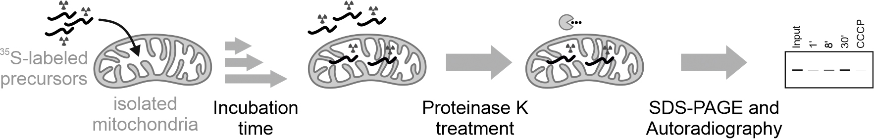
Overview of the assay to monitor protein import into mitochondria isolated from human cells
Background
Mitochondrial proteins perform critical functions in energy conversation, heat production, lipid and iron metabolism, signaling, and cell death pathways (Vafai and Mootha, 2012; Spinelli and Haigis, 2018; Habich et al., 2019b; Pfanner et al., 2019). Human mitochondria contain approximately 1500 different proteins, but only 13 are translated in the matrix on mitochondrial ribosomes. Thus, the vast majority of proteins are synthesized as precursors on cytosolic ribosomes and then imported into mitochondria. Different import pathways have been identified that direct proteins to their correct submitochondrial localization (Hartl et al., 1989; Chacinska et al., 2009; Schmidt et al., 2010; MacPherson and Tokatlidis, 2017; Endo and Tamura, 2018; Hansen and Herrmann, 2019). Most of these pathways have been characterized using a handful of model substrates. However, it becomes increasingly clear that many proteins have different dependencies on import machinery components and that the intrinsic protein properties that drive import can vary. In vitro reconstituted systems using isolated mitochondria are powerful tools that allow rapid analysis of mitochondrial protein import. These assays allow rapid and parallel analysis of protein import and its determinants. In contrast to assays in intact cells, cytosolic processes – including proteasomal degradation, differing protein transcription/translation rates, competition with other precursors, and contributions of specific chaperone systems – can be excluded in import assays using isolated mitochondria.
Most described import experiments have been performed in mitochondria isolated from S. cerevisiae (Mokranjac and Neupert, 2007; Weckbecker and Herrmann, 2013; Tang and Tang, 2015), mainly because yeast genetics allowed detailed mechanistic analysis and insight into import pathways. We adapted the protocol for monitoring protein import into mitochondria enriched from human tissue culture cells. The combination with CRISPR-Cas9 mediated gene editing (knock-outs and knock-ins), siRNA-mediated protein depletion, and stable cell lines overexpressing components of the human import machinery allow a detailed investigation of human mitochondrial protein import. This enables the investigation of the differences between human and yeast mitochondrial import as well as the analysis of human patient mutations.
Materials and Reagents
Cultivation of HEK293 cells
Cell culture dishes 100 × 20 mm (Sarstedt, catalog number: 83.3902)
Cell culture dishes 150 × 20 mm (Sarstedt, catalog number: 83.3903)
Autoclaved disposable glass Pasteur pipets without cotton pad (VWR, catalog number: HECH40567001)
Serological pipettes: Sterile individually packed,
2 ml pipettes (Sarstedt, catalog number: 83.3903)
5 ml pipettes (Sarstedt, catalog number: 86.1252.001)
10 ml pipettes (Sarstedt, catalog number: 86.1254.001)
25 ml pipettes (Sarstedt, catalog number: 86.1685.001)
Flp-In T-REx HEK293 cells (Human embryonic kidney 293, Invitrogen, catalog number: R78007)
Dulbecco’s modified Eagle medium (DMEM) medium-complete (see Recipes)
10% Fetal calf serum (FCS, Sigma-Aldrich, catalog number: F0804) and
1% penicillin/streptomycin (P/S, Sigma-Aldrich, catalog number: P0781-100ML)
DMEM high glucose (Thermo Fisher, catalog number: 41965-062)
Dulbecco's Phosphate Buffered Saline (PBS) (see Recipes)
PBS powder (Sigma-Aldrich, catalog number: D5652)
Trypsin-EDTA solution (see Recipes)
10× Trypsin-EDTA solution (Sigma-Aldrich, catalog number: T4174)
Isolation of mitochondria from HEK293 cells
Cell scraper 16 mm (Sarstedt, catalog number: 831832)
15 ml Falcon tubes (VWR, catalog number: 734-0451)
3 × 150 mm confluent dishes plated with HEK293 cells
Note: Cells are seeded 3-4 days prior to the mitochondrial isolation. Cells are grown to 90% confluency. See Note 2.
CompleteTM Protease Inhibitor Cocktail EDTA-free tablets (Roche, catalog number: 11873580001). Add freshly before use. Aliquots can be stored at -20°C
PBS, store at 4°C before use
Bradford Reagent ROTI® Quant (Roth, catalog number: K015.1)
1× buffer M (see Recipes)
Mannitol (Merck, catalog number: 1.05983)
Sucrose (Sigma-Aldrich, catalog number: S0389)
HEPES-KOH, pH 7.4 (VWR, catalog number: 441487)
EGTA-KOH, pH 7.4 (Roth, catalog number: 3054.1)
Synthesis of [35S]-labeled precursor
TNT® Quick Coupled Transcription/Translation System SP6 Promoter (Promega, catalog number: L2080). See Note 3.
EasyTagTM EXPRESS 35S Protein Labeling Mix, [35S]-, 7 mCi, 1175 Ci/mmol (PerkinElmer, catalog number: NEG772007MC). See Note 4.
DNA template (1 µg ml-1) containing the gene of the mitochondrial precursor inserted downstream of the SP6 promoter. Plasmid vector: pGEM-3/4 (Promega). Store at -20°C.
Nuclease-free double-distilled water
200 mM L-methionine (Merck, catalog number: 1057070100) in nuclease-free double-distilled water. Store at 4°C
50 mM ethylene diamine tetra acidic acid (EDTA) disodium salt 2-hydrate, pH 8 (AppliChem, catalog number: A2937) in nuclease-free double-distilled water
1× sample buffer (see Recipes)
Tris(hydroxymethyl)aminomethane (Tris, VWR, catalog number: 0497)
Glycerol (Sigma-Aldrich, catalog number: G7757)
Bromophenol blue (Roth, catalog number: A512.1)
Dithiothreitol (DTT, AppliChem, catalog number: A1101)
Import of [35S]-labeled precursors into isolated mitochondria
Blotting membrane, nitrocellulose, AmershamTM Protran® (VWR, catalog number: 10600001), 0.2 µm pore size
20 mM Carbonyl cyanide 3-chlorophenylhydrazone (CCCP, Sigma-Aldrich, catalog number: C2759)
200 mM phenylmethylsulfonyl fluoride (PMSF, AppliChem, catalog number: A0999.0100)
Crushed ice
Triton X-100 (AppliChem, catalog number: A1388)
1× buffer M (see Recipes)
20 µg/import reaction mitochondria (see: Recipes)
20 µg ml-1 peqGOLD proteinase K (see Recipes)
1× sample buffer (see Recipes)
SEH buffer (see Recipes)
Equipment
Cultivation of HEK293 cells
Laminar flow hood class II (ENVAIR eco)
CO2 incubator
Vacuum pump
Microscope
Centrifuge (e.g., Eppendorf, model: 5702R)
Equipped cell culture laboratory containing, e.g., a laminar flow hood class II (ENVAIR eco), a CO2 incubator for cell cultivation set at 37°C and 5% CO2, a vacuum pump for removal of medium using sterile glass Pasteur pipets, a microscope, and a cell counter and a cooling centrifuge.
Isolation of mitochondria from HEK293 cells
Electric stirrer (Heidolph RZR 2020)
Potter S homogenizer (15 ml) with tight-fitting PTFE (polytetrafluorethylen) plungers (Sartorius, catalog number: 8542406)
The range of clearance (i.e., the average distance between the pestle and the cylinder walls) optimal for mitochondria isolation is thereby from 0.045-0.065 mm.
Centrifuge (Eppendorf, model: 5702R)
Centrifuge (Eppendorf, model: 5417R)
Vacuum pump (KNF laboport minipump) for removal of medium using sterile glass Pasteur pipets.
[35S]-labeled precursor synthesis
Hypoxic Glove Box and Cabinet (Coy Laboratory Products, “Coy 1 Person O2 Control Glove Box”, catalog number: 031615); not necessary if precursor protein is not prone to air oxidation (e.g., does not contain or contains only few cysteine residues).
Import of [35S]-labeled precursors into isolated mitochondria
ThermoMixer Eppendorf (F1.5, #EP5384000012)
Centrifuge (Eppendorf, model: 5417R)
System for SDS-PAGE electrophoresis and (semidry) blotting (Bio-Rad, MiniPROTEAN Tetra Cell, catalog number: 1658000)
Imaging System Typhoon FLA 9500 (GE Healthcare, catalog number: 9351515)
Image Eraser FLA (GE Healthcare, catalog number: 12999750)
Software
ImageQuant TL 8.1 (GE Healthcare Life Science).
Procedure
文章信息
版权信息
© 2021 The Authors; exclusive licensee Bio-protocol LLC.
如何引用
Murschall, L. M., Peker, E., MacVicar, T., Langer, T. and Riemer, J. (2021). Protein Import Assay into Mitochondria Isolated from Human Cells. Bio-protocol 11(12): e4057. DOI: 10.21769/BioProtoc.4057.
分类
生物化学 > 蛋白质 > 翻译后修饰
细胞生物学 > 细胞器分离 > 线粒体
您对这篇实验方法有问题吗?
在此处发布您的问题,我们将邀请本文作者来回答。同时,我们会将您的问题发布到Bio-protocol Exchange,以便寻求社区成员的帮助。