

Quantitation of Secretory Granule Size in Drosophila Larval Salivary Glands
果蝇幼虫唾液腺分泌颗粒大小的定量研究
发布: 2021年06月05日第11卷第11期 DOI: 10.21769/BioProtoc.4039 浏览次数: 3879
评审: Gal HaimovichSteve JeanPradeep Kumar Bhaskar
参见作者原研究论文
The authors used this protocol in:
Mar 2020


Abstract
Maturation of secretory granules is a crucial process that ensures the bioactivity of cargo proteins undergoing regulated secretion. In Drosophila melanogaster, the larval salivary glands produce secretory granules that are up to four-fold larger in cross-sectional area after maturation. Therefore, we developed a live imaging microscopy approach to quantitate the size of secretory granules with a view to identifying genes involved in their maturation. Here, we describe the procedures of larval salivary gland dissection and sample preparation for live imaging with a fluorescence confocal microscope. Furthermore, we describe the workflow for measuring the size of secretory granules by cross-sectional surface area and statistical analysis. Our live imaging microscopy method provides a reliable read-out for the status of secretory granule maturation in Drosophila larval salivary glands.
Keywords: Drosophila (果蝇)Background
Regulated secretion is a process during which biologically active molecules such as hormones, digestive enzymes, and mucus are secreted from specialized secretory cells in a coordinated manner. Hence, regulated secretion is critical for maintaining physiological homeostasis in animals. Examples include the release of insulin after a meal, the release of mucin in response to pathogenic microorganisms, and the release of sweat during elevated body temperature. These biologically active molecules are produced by endocrine or exocrine cells and stored in long-lasting secretory organelles termed secretory granules.
Biogenesis of secretory granules begins at the trans-Golgi network, where cargoes of secretory granules aggregate and bud off as immature secretory granules (Tooze, 1991 and 1998; Borgonovo et al., 2006). Immature secretory granules then undergo a maturation process to become fully functional and competent for secretion. The maturation process includes homotypic fusion of immature secretory granules, removal of unwanted materials, and processing of cargoes (Tooze, 1991; Arvan and Castle, 1998). Failure of secretory granules to mature can lead to reduced bioactivity of their cargoes. For example, most hormones enter immature secretory granules as inactive prohormones. During maturation, the lumen of secretory granules is acidified, and prohormone convertases cleave prohormones into biologically active hormones (Moore et al., 2002). Therefore, failed granule maturation can have physiological consequences, leading to reduced activity or inefficient secretion of cargo proteins.
Although secretory granule maturation is a critical step in regulated secretion, assays for secretory granule maturation are not simple. Transmission electron microscopy is one of the standard methods to assay secretory granule maturation. Secretory granules are electron dense in electron micrographs (Nitsch and Rinne, 1981; Tatsuoka and Reese, 1989). Reduced secretory granule maturation is often correlated with a reduction in electron density or in secretory granule number (Edwards et al., 2009; Cao et al., 2013; Du et al., 2016; Emperador-Melero et al., 2018; Hummer et al., 2017; Rao et al., 2020). If antibodies are available, a decrease in hormone secretion or an increased ratio of prohormone to hormone can be measured in blood plasma or cell culture media by western blotting and densitometry or ELISA following stimulation (Cao et al., 2013; Du et al., 2016; Hummer et al., 2017). Immunofluorescence using a combination of anti-prohormone antibodies and secretory granule markers can also reveal defects in hormone processing, as indicated by an increase in intensity of the prohormone signal (Bogan et al., 2012; Cao et al., 2013).
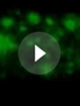
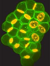
The Drosophila larval salivary gland is a powerful genetic model for studying secretory granule biogenesis (Biyasheva et al., 2001; Burgess et al., 2011 and 2012; Torres et al., 2014; Csizmadia et al., 2018; Ma et al., 2020; Neuman et al., 2020). The larval salivary glands start producing glue protein-containing secretory granules 24 h after entry into the third instar larval stage. Immature secretory granules then mature over the next 18 h and are secreted all at once in response to a pulse of the hormone ecdysone (Biyasheva et al., 2001; Burgess et al., 2011). The secreted glue proteins adhere pupal cases onto a solid surface during metamorphosis. Size differences between immature and mature secretory granules can be 2- to 4-fold in cross-sectional surface area (5 μm2 vs. 10-25 μm2) (Ma et al., 2020). Thus, the Drosophila larval salivary gland is an excellent system to identify genes required for secretory granule maturation.
The Andres laboratory previously generated transgenic lines expressing one of the glue proteins (Sgs3) tagged with GFP or DsRed under the control of its endogenous promoter (Biyasheva et al., 2001; Costantino et al., 2008). Using these lines, secretory granules can be visualized by confocal microscopy. Our laboratory has used these transgenic lines in combination with a salivary gland-specific Gal4 driver and UAS-controlled RNAi transgenic lines to identify genes needed for secretory granule maturation. Here, we provide a detailed protocol for visualizing secretory granules via live imaging with a spinning-disc confocal microscope. Acquired data are analyzed with the imaging software Volocity 6.3 to quantitate secretory granule size. Secretory granule size distribution is then used as a readout for secretory granule maturation in the Drosophila larval salivary gland.
Materials and Reagents
Microscope slides 75 × 25 × 1 mm (VWR, catalog number: 16004-368)
Glass coverslips 18 × 18 mm No. 1.5 (VWR, catalog number: 48366-205)
Self-adhesive reinforcement labels (Avery, catalog number: 32203)
Syringe 5 ml (BD, catalog number: 309603)
Needle 18 G or lower (BD, catalog number: 305195)
Dissection needle (Fisher Scientific, catalog number: 13820024)
Petri dishes 100 × 15 mm (VWR, catalog number: 25384-088)
Petri dishes 35 × 10 mm (Corning, catalog number: 351008)
P{w+, Sgs3-GFP} (Bloomington Drosophila Stock Center, catalog number: 5884, 5885) or P{w+, Sgs3-DsRed} (Andres lab, University of Nevada, Las Vegas, USA) 3rd instar larva
High-vacuum M grease 100 g (Apiezon, Sigma, catalog number: Z273589)
Pipette tips (P200)
SYLGARDTM 184 Silicone Elastomer Kit 0.5 kg (Dow Chemical, Sigma, catalog number: 4019862)
KCl (Sigma, catalog number: P3911-500G)
NaCl (Bio Basic, catalog number: CA99501-558)
CaCl2·2H2O (Sigma, catalog number: C-5080-500G)
Tris-HCl (VWR, catalog number: 0234-1KG)
Drosophila Ringer’s solution (see Recipe 1)
Silicone dissection plate (see Recipe 2)
Equipment
Dumont #5 fine forceps (Fine Science Tools, catalog number: 11251-20)
Stereomicroscope (Leica Microsystems, model: Leica MZ6)
Spinning-disc confocal coupled with an Olympus IX81 microscope (Quorum Technologies, Puslinch, Ontario, Canada)
Software
Volocity 6.3 (Quorum Technologies, Puslinch, ON, Canada)
Adobe Creative Cloud Photoshop (Adobe, San Jose, CA, USA)
Microsoft Office 365 Excel (Microsoft, Redmond, WA, USA)
Procedure
文章信息
版权信息
© 2021 The Authors; exclusive licensee Bio-protocol LLC.
如何引用
Readers should cite both the Bio-protocol article and the original research article where this protocol was used:
- Ma, C. J. and Brill, J. A. (2021). Quantitation of Secretory Granule Size in Drosophila Larval Salivary Glands. Bio-protocol 11(11): e4039. DOI: 10.21769/BioProtoc.4039.
- Ma, C. J., Yang, Y., Kim, T., Chen, C. H., Polevoy, G., Vissa, M., Burgess, J. and Brill, J. A. (2020). An early endosome-derived retrograde trafficking pathway promotes secretory granule maturation.J Cell Biol 219 (3): e201808017.
分类
发育生物学 > 形态建成 > 器官形成
细胞生物学 > 细胞成像 > 活细胞成像
细胞生物学 > 细胞分离和培养 > 器官培养
您对这篇实验方法有问题吗?
在此处发布您的问题,我们将邀请本文作者来回答。同时,我们会将您的问题发布到Bio-protocol Exchange,以便寻求社区成员的帮助。