

Intracellular IRF5 Dimerization Assay
细胞内IRF5二聚分析
发布: 2021年05月20日第11卷第10期 DOI: 10.21769/BioProtoc.4021 浏览次数: 6889
评审: Martin V KolevAnonymous reviewer(s)

参见作者原研究论文
The authors used this protocol in:

May 2020
Advertisement


Abstract
The intracellular interferon regulatory factor 5 (IRF5) dimerization assay is a technique designed to measure molecular interaction(s) with endogenous IRF5. Here, we present two methods that detect endogenous IRF5 homodimerization and interaction of endogenous IR5 with cell penetrating peptide (CPP) inhibitors. Briefly, to detect endogenous IRF5 dimers, THP-1 cells are incubated in the presence or absence of the IRF5-targeted CPP (IRF5-CPP) inhibitor for 30 min then the cells are stimulated with R848 for 1 h. Cell lysates are separated by native-polyacrylamide gel electrophoresis (PAGE) and IRF5 dimers are detected by immunoblotting with IRF5 antibodies. To detect endogenous interactions between IRF5 and FITC-labeled IRF5-CPP, an in-cell fluorescence resonance energy transfer (FRET) assay is used. In this assay, THP-1 cells are left untreated or treated with FITC-IRF5-CPP conjugated inhibitors for 1 h. Next, cells are fixed, permeabilized, and stained with anti-IRF5 and TRITC-conjugated secondary antibodies. Transfer of fluorescence can be measured and calculated as FRET units. These methods provide rapid and accurate assays to detect IRF5 molecular interactions.
Keywords: IRF5 (细胞内干扰素调节因子5)Background
Interferon regulatory factor 5 (IRF5) is a transcription factor that regulates pathogen-induced innate and acquired immune responses downstream of Toll-like receptor (TLR), retinoic acid-inducible gene I (RIG-I), melanoma differentiation-associated gene 5 (MDA5), and B cell receptor (BCR) (De et al., 2017; Thompson et al., 2018; Banga et al., 2020). IRF5 has been implicated in the pathogenesis of systemic lupus erythematosus due to its role in regulating the expression of proinflammatory cytokines such as IFN-α, IL-6, TNF-α, and IL-12 and pathogenic autoantibody production (Song et al., 2020). In an unstimulated condition, IRF5 is generally localized in the cytoplasm as a monomer (Thompson et al., 2018). Activation of the above receptors triggers cellular signaling cascades. IRF5 undergoes post-translational modification, which eventually leads to homodimerization, a critical event prior to nuclear translocation (Thompson et al., 2018). Here, we describe two methods designed for detecting IRF5 molecular interactions.
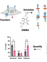
A native-PAGE method is used to detect endogenous IRF5 homodimers. THP-1 cells are incubated with or without (1 and 10 μM) IRF5-CPP inhibitors for 30 min, and subsequently stimulated with 1 μM of R848 (a TLR7 ligand) or left unstimulated for 1 h. Next, cell lysates are run on native polyacrylamide gel electrophoresis (PAGE) and IRF5 dimerization is detected by immunoblotting with IRF5 and HRP-conjugated secondary antibodies. The intracellular fluorescence resonance energy transfer (FRET) assay is used to detect binding of endogenous IRF5 to FITC-IRF5-CPP conjugated inhibitors through a FRET signal (Banga et al., 2020; Song et al., 2020). FRET is a technique designed for detecting molecular interaction in which the excited molecule (the donor) transfers non-radiative energy to another molecule (the acceptor) within a distance of ~1-10 nm (Doucey et al., 2003; Ujlaky-Nagy et al., 2018; Banga et al., 2020). THP-1 cells are incubated with or without (1 and 10 μM) IRF5-CPP inhibitors for 1 h. The untreated and FITC-IRF5-CPP- treated THP-1 cells are then fixed, permeabilized, and stained with anti-IRF5, anti-IRF3, or anti-IRF7 (other IRF family members) antibodies and TRITC secondary antibodies. Cell-associated fluorescence is measured on a BioTek Synergy Neo2 at 525 nm upon excitation at 488 nm (E1), at 600 nm upon excitation at 540 nm (E2), and at 600 nm upon excitation at 488 nm (E3). The transfer of fluorescence is calculated as FRET units as follows: FRET unit = (E3both − E3none) − ([E3TRITC − E3none) × (E2both/E2TRITC]) − ([E3FITC − E3none] × [E1both/E1FITC]) (Doucey et al., 2003; Banga et al., 2020; Song et al., 2020). The different fluorescence values (E) were measured on unlabeled cells (Enone) or cells labeled with FITC (EFITC) and TRITC (ETRITC) (Banga et al., 2020; Song et al., 2020).
These techniques can be broadly applied to evaluate intracellular molecular interactions in living cells through pharmacological and molecular studies. They provide a rapid and reliable method of screening molecular interactions, which require standard equipment that are readily available in almost every laboratory.
Part I. Native-PAGE (Polyacrylamide Gel Electrophoresis)
Materials and Reagents
THP-1 cells (ATCC, catalog number: TIB-202)
RPMI-1640 medium (Gibco, catalog number: 11875093)
Fetal bovine serum (Gibco, catalog number: 16000044)
Penicillin-streptomycin (Gibco by Life Technologies, catalog number: 15140122)
IRF5 cell penetrating peptides (IRF5-CPP) and FITC-IRF5-CPP inhibitors (Hoffman-LaRoche, Patent number WO2014001229A2)
Resiquimod (R848) (Millipore Sigma, catalog number: SML0196)
Sodium deoxycholate (DOC) (Thermo Fisher, catalog number: 89904)
DC protein assay (Bio-Rad, catalog number: 5000111)
BCA protein assay (Thermo Fisher, catalog number: 23227)
Anti-IRF3, rabbit monoclonal antibody (Abcam, catalog number: ab76409)
Anti-IRF5 (Cell Signaling Tech, catalog number: 3257, rabbit polyclonal antibody or catalog number: 13496, rabbit monoclonal antibody)
Anti-IRF7, rabbit polyclonal antibody (Cell Signaling Tech, catalog number: 4920)
Horseradish peroxidase (HRP)–conjugated anti-β-actin, rabbit monoclonal antibody (Cell Signaling Tech, catalog number: 5125)
Anti-rabbit IgG HRP-conjugated secondary antibody (Cell Signaling Tech, catalog number: 7074)
NP40 cell lysis buffer (Invitrogen, catalog number: FNN0021)
Phenylmethylsulfonyl fluoride (PMSF) (Millipore Sigma, catalog number: 10837091001)
Protease inhibitor (Millipore Sigma, catalog number: P-2714)
Native gel running buffer, Tris-glycine buffer 10× concentrate, pH 8.3 (Millipore Sigma, catalog number: T4904-1L)
Glycerol (Millipore Sigma, catalog number: G9012)
Immobilon-P 0.45 μm PVDF membrane (Millipore, catalog number: IPVH00010)
Bovine serum albumin (BSA) (Millipore Sigma catalog number: 05470-5G)
Nonfat-dried milk bovine (Millipore Sigma, catalog number: M7409-1BTL)
Bromophenol blue (Millipore Sigma, catalog number: B5525)
30% acrylamide/Bis solution, 37.5:1, 500 ml (Bio-Rad, catalog number: 161-0158)
Resolving gel buffer, 1.5 M Tris-HCl, pH 8.8, 1 L (Bio-Rad, catalog number: 161-0798)
Stacking gel buffer, 0.5 M Tris-HCl, pH 6.8, 1 L (Bio-Rad, catalog number: 161-0799)
SDS solution, 10% (w/v), 250 ml (Bio-Rad, catalog number: 161-0416)
Pierce 20× TBS Tween 20 buffer (TBST) (Thermo Fisher, catalog number: 28360)
Western bright ECL HRP substrate (Advansta, catalog number: K-12045-C20)
Precision plus protein standard (Bio-Rad, catalog number: 161-0374) in which high molecular weight bands can be detected on native gel
Equipment
ChemiDoc MP Imaging System (Bio-Rad, catalog number: 17001402)
Mini-Transblot Cell and PowerPac Basic Power Supply (Bio-Rad, catalog number: 1703989)
Mini-Protean Tetra Vertical Electrophoresis Cell for Mini Precast Gels (Bio-Rad, catalog number: 1658005)
Procedure
文章信息
版权信息
© 2021 The Authors; exclusive licensee Bio-protocol LLC.
如何引用
Readers should cite both the Bio-protocol article and the original research article where this protocol was used:
- Sherman, C. D. and Barnes, B. J. (2021). Intracellular IRF5 Dimerization Assay. Bio-protocol 11(10): e4021. DOI: 10.21769/BioProtoc.4021.
- Banga, J., Srinivasan, D., Sun, C. C., Thompson, C. D., Milletti, F., Huang, K. S., Hamilton, S., Song, S., Hoffman, A. F., Qin, Y. G., Matta, B., LaPan, M., Guo, Q., Lu, G., Li, D., Qian, H., Bolin, D. R., Liang, L., Wartchow, C., Qiu, J., Downing, M., Narula, S., Fotouhi, N., DeMartino, J. A., Tan, S. L., Chen, G. and Barnes, B. J. (2020). Inhibition of IRF5 cellular activity with cell-penetrating peptides that target homodimerization.Sci Adv 6(20): eaay1057.
分类
生物化学 > 蛋白质 > 相互作用 > 蛋白质-蛋白质相互作用
生物化学 > 蛋白质 > 相互作用 > 蛋白质-配体相互作用
生物化学 > 蛋白质 > 荧光
您对这篇实验方法有问题吗?
在此处发布您的问题,我们将邀请本文作者来回答。同时,我们会将您的问题发布到Bio-protocol Exchange,以便寻求社区成员的帮助。