

ATAC-Seq-based Identification of Extrachromosomal Circular DNA in Mammalian Cells and Its Validation Using Inverse PCR and FISH
基于ATAC-Seq的哺乳动物细胞染色体外环状DNA鉴定及其反向PCR和FISH验证
(*contributed equally to this work) 发布: 2021年05月05日第11卷第9期 DOI: 10.21769/BioProtoc.4003 浏览次数: 9027
评审: Manasi K. MayekarAnonymous reviewer(s)
参见作者原研究论文
The authors used this protocol in:

May 2020
Advertisement


Abstract
Recent studies from multiple labs including ours have demonstrated the importance of extrachromosomal circular DNA (eccDNA) from yeast to humans (Shibata et al., 2012; Dillon et al., 2015; Møller et al., 2016; Kumar et al., 2017; Turner et al., 2017; Kim et al., 2020). More recently, it has been found that cancer cells obtain a selective advantage by amplifying oncogenes on eccDNA, which drives genomic instability (Wu et al., 2019; Kim et al., 2020). Previously, we have purified circular DNA and enriched the population using rolling circle amplification followed by high-throughput sequencing for the identification of eccDNA based on the unique junctional sequence. However, eccDNA identification by rolling circle amplification is biased toward small circles. Here, we report a rolling circle-independent method to detect eccDNA in human cancer cells. We demonstrate a sensitive and robust step-by-step workflow for finding novel eccDNAs using ATAC-seq (Assay for Transposase-Accessible Chromatin using sequencing) combined with a Circle_finder bioinformatics algorithm to predict the eccDNAs, followed by its validation using two independent methods, inverse PCR and metaphase FISH (Fluorescence in situ Hybridization).
Keywords: Circular DNA (环状DNA)Background
Extrachromosomal circular DNAs (eccDNAs) are unique DNA molecules that carry genetic information in addition to the chromosomal DNAs. These eccDNAs have been found in different organisms from yeast to humans (Shibata et al., 2012; Dillon et al., 2015; Møller et al., 2016; Kumar et al., 2017; Turner et al., 2017; Kim et al., 2020). The length of eccDNAs ranges from small (less than 1kb, also called microDNAs) to large (megabase-long). While the small eccDNAs with micro homology ends may promote genetic heterogeneity (Shibata et al., 2012) or produce short RNAs if transcribed (Paulsen et al., 2019), the long eccDNAs may harbor complete genes and regulatory elements such as enhancers (Morton et al., 2019; Wu et al., 2019; Koche et al., 2020). Emerging evidence suggests that eccDNAs could play underappreciated roles in regulating gene expression and genome instability, which ultimately contributes to the selective advantage of cells (Gresham et al. 2010, Koo et al., 2018; Hull et al., 2019). In particular, oncogene-carrying eccDNAs are highly amplified in human cancers and correlate with open chromatin, increased oncogene expression, and chromosome structural rearrangement, in addition to being associated with poor outcomes (Wu et al., 2019; Kim et al., 2020; Koche et al., 2020). Uncovering eccDNAs in the circulation also makes them prospective targets for diagnostic purposes (Kumar et al., 2017; Sin et al., 2020).
The growing research on eccDNAs calls for tool development for eccDNA discovery. Historically, eccDNAs of various sizes were detected by karyotyping, electron microscopy, Southern blotting, and 2-D gel electrophoresis (reviewed in Paulsen et al., 2018). More recently, various high-throughput sequencing (HTS) technologies have been exploited to facilitate the discovery of new eccDNAs (Shibata et al., 2012; Møller et al., 2015; Kim et al., 2020). The basic idea for eccDNA detection via HTS methods is based on their distinctive circular feature – eccDNAs of high confidence can be identified from paired-end reads that (1) could not map as inward pairs on the linear genome, and (2) contain the unique circular junctional sequence that represents the chromosome breakage/ligation point. A unique junctional sequence (shown as “E-A” in Figure 1A) that is not present in the normal reference genome could be formed through ligation of the two ends of a linear DNA, thus creating the circular DNA. However, the majority of eccDNA sequencing pipelines utilize multiple displacement amplification (MDA), an efficient method to amplify small amounts of DNA via rolling circle amplification, which would preferentially amplify short circles. Therefore, we sought to develop an MDA-independent pipeline that incorporates several additional validation assays.
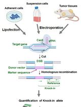
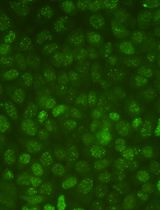
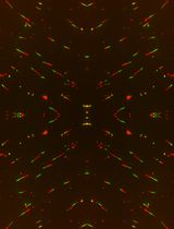
Recently, we demonstrated a robust workflow to detect and validate new eccDNAs from human cancer cell cultures (Kumar et al., 2020). Specifically, ATAC-seq (Assay for Transposase-Accessible Chromatin using sequencing) and a Circle_finder algorithm was employed for new eccDNA prediction. ATAC-seq, first developed in 2013, utilizes engineered Tn5 transposase to cut open chromatin regions (including eccDNAs that are less chromatinized) and insert transposase-associated adapter DNAs (Buenrostro et al., 2013 and 2015). The Circle-finder algorithm (refer to Software for link access) predicts eccDNAs from paired-end sequencing based on: (1) the presence of split reads (one read maps to two sites in the genome); (2) the two fragments on the split read maps on the same chromosome and same strand; and (3) the continuous read maps between the two fragments on the split read and on the opposite strand to the split read. Predicted eccDNAs can be evaluated by two independent validation assays (Figure 1). Inverse PCR (Figure 1B) will specifically amplify eccDNAs with a primer pair that spans the unique junctional sequence (shown as “E-A”); such a primer pair faces outward on genomic DNA and results in no amplification. Alternatively, eccDNA can be visually confirmed by metaphase FISH (Figure 1C), which can detect both genomic DNA signals overlapping with main chromosomes and signals from eccDNAs that do not overlap with chromosomes.

Figure 1. Overview of eccDNA identification and validation by ATAC-seq, inverse PCR, and metaphase FISH
Materials and Reagents
50 ml Falcon conical tubes (Fisher Scientific, catalog number: 1443222)
15 ml Falcon conical tubes (Fisher Scientific, catalog number: 1495949B)
1.5 ml DNA LoBind tubes (Eppendorf, catalog number: 022431021)
1 ml pipette tip
100-200 μl pipette tip
100 mm Petri dish
0.2 μm filter
Microscope glass slides (Fisher Scientific, catalog number: 4951F-001)
22 mm × 50 mm cover glass (Fisher Scientific, catalog number: 12-545E)
Parafilm (Thermo Fisher Scientific, catalog number: S37440)
Aluminum foil (Thermo Fisher Scientific, catalog number: 14-648-236)
Mammalian cells in culture. In this protocol, we used the ovarian cancer cell line, OVCAR8, and the prostate cancer cell line, C4-2B, cultured in RPMI medium (Corning, catalog number: 10-040-CV) supplemented with 10% fetal bovine serum (Thermo Fisher Scientific, catalog number: 26140079) and 1% penicillin-streptomycin (Thermo Fisher Scientific, catalog number: 15-140-122)
SybrGold dye (Invitrogen, catalog number: S11494)
0.5% trypsin-EDTA (Thermo Fisher Scientific, catalog number: 15400054)
UltraPure DNase/RNase-free distilled water (Thermo Fisher Scientific, catalog number: 10977015)
1 M Tris-HCl, pH 7.5 (Thermo Fisher Scientific, catalog number: 15567027, store at 4°C, shelf life: 6 months)
5 M NaCl solution (Thermo Fisher Scientific, catalog number: AM9760G, store at room temperature)
1 M MgCl2 solution (Thermo Fisher Scientific, catalog number: AM9530G, store at room temperature)
Dulbecco’s phosphate-buffered saline or DPBS, no calcium, no magnesium (Thermo Fisher/Gibco, catalog number: 14190144, store at 4°C, shelf life: 36 months)
10% Nonidet P40 substitute (Millipore/Sigma-Aldrich, catalog number: 11332473001, store at 4°C, keep protected from light, shelf life: 24 months)
10% (w/v) Tween-20 (Millipore/Sigma-Aldrich, catalog number: 11332465001, store at 4°C under inert gas and keep protected from light, shelf life: 24 months)
20 mg/ml digitonin in DMSO (Promega, catalog number: G9411, store in -20 °C)
DNA Clean & Concentrator Kit (ZYMO, catalog number: D4033)
Nextera DNA Sample Preparation Kit (Illumina, catalog number: FC-121-1030, store at -20°C)
Note: This kit has been discontinued and can be purchased separately: Tagmentation DNA Enzyme/TDE (Illumina, catalog number: 15027865) and Tagmentation DNA Buffer/TDB (Illumina, catalog number: 15027866).Nextera Index Kit, 24 indexes (Illumina, catalog number: 15055289, store at -20°C)
NEBNext High-Fidelity 2× PCR Master Mix (New England Biolabs, catalog number: M0541, store at -20°C)
Phosphate-buffered saline, pH 7.4 (Thermo Fisher Scientific, catalog number: 10010023)
Qiagen HiSpeed Plasmid Midi Kit (Qiagen, catalog number: 12643)
Isopropanol (Fisher Chemical, catalog number: A516-500)
Ethanol (Thermo Fisher Scientific, catalog number: A4094)
Glycogen (Thermo Fisher Scientific, catalog number: AM9510)
Plasmid-safe ATP-dependent DNase (Lucigen, catalog number: E3101K)
QIAquick PCR Purification Kit (Qiagen, catalog number: 28104)
KOD Hot-Start DNA Polymerase (Millipore/Sigma-Aldrich, catalog number: 71086)
Thymidine (Millipore/Sigma-Aldrich, catalog number: T1895)
10 mg/ml KaryoMax Colcemid solution in PBS (Thermo Fisher Scientific, catalog number: 15212012)
Potassium chloride (Millipore/Sigma-Aldrich, catalog number: P9541)
Formamide (Millipore/Sigma-Aldrich, catalog number: 47670)
Sodium chloride (Thermo Fisher Scientific, catalog number: BP358)
Sodium citrate (Millipore/Sigma-Aldrich, catalog number: W302600)
BAC FISH Probe label with 5-fluorescein (Empire Genomics, catalog number: RP11-732I3 and RP11-765O11)
Rubber cement (Elmer’s Rubber Cement, catalog number: EPIE904)
Nonidet P-40 (Sigma, catalog number: I8896)
Dextran sulfate (Thermo Fisher Scientific, catalog number: BP1585)
VectaShield Mounting Medium with DAPI (Vector Laboratories, catalog number: H-1200-10)
Nail polish (OPI Nail Lacquer)
Nikon immersion oil for the confocal microscope (Thermo Fisher Scientific, catalog number: 12-624-66A)
1% (10 mg/ml) digitonin (see Recipes, store at -20°C as aliquotes, stable for 6 months)
ATAC-Resuspension Buffer (see Recipes)
ATAC-Lysis Buffer (see Recipes)
ATAC-Wash Buffer (see Recipes)
ATAC-Reaction Mastermix (see Recipes)
100 mM thymidine solution (see Recipes)
75 mM KCl Hypotonic Solution (see Recipes)
Carnoy’s Fixative Solution (see Recipes)
20× Saline-Sodium-Citrate (SSC) buffer (see Recipes)
Hybridization Buffer (see Recipes)
FISH Denaturation Buffer (see Recipes)
FISH Wash Buffer 1 (see Recipes)
FISH Wash Buffer 2 (see Recipes)
Equipment
Cell culture incubator
Tissue culture hood
Tabletop microcentrifuge (Eppendorf, model: 5424)
Thermomixer (Thermo Scientific, catalog number: 13687711)
PCR machine with heated lid (Eppendorf, model: Mastercycler Pro)
Tabletop centrifuge (Eppendorf, model: 5804)
Water bath (Thermo Fisher Scientific, Isotemp)
Coplin jar (Local Company)
Hybridization chamber (Thermo Fisher Scientific, Isotemp)
Chemical fume hood (Bellco Glass Inc.)
Brightfield microscope (Olympus)
Confocal microscope (Nikon, model: Ti-E eclipse series)
Computer with enough data storage capacity up to TB
Software
Circle_finder (github, https://github.com/pk7zuva/Circle_finder/blob/master/circle_finder-pipeline-bwa-mem-samblaster.sh); pre-requisite installation to run Circle_finder: bedtools (https://github.com/arq5x/bedtools2), samtools (http://samtools.sourceforge.net), parallel (https://www.gnu.org/software/parallel/), bwa (https://github.com/lh3/bwa), samblaster (https://github.com/GregoryFaust/samblaster)
Cutadapt (https://cutadapt.readthedocs.io/en/stable/)
AR Elements Software (Nikon, Japan)
ImageJ (NIH, USA)
Procedure
文章信息
版权信息
© 2021 The Authors; exclusive licensee Bio-protocol LLC.
如何引用
Readers should cite both the Bio-protocol article and the original research article where this protocol was used:
- Su, Z., Saha, S., Paulsen, T., Kumar, P. and Dutta, A. (2021). ATAC-Seq-based Identification of Extrachromosomal Circular DNA in Mammalian Cells and Its Validation Using Inverse PCR and FISH. Bio-protocol 11(9): e4003. DOI: 10.21769/BioProtoc.4003.
- Kumar, P., Kiran, S., Saha, S., Su, Z., Paulsen, T., Chatrath, A., Shibata, Y., Shibata, E. and Dutta, A. (2020). ATAC-seq identifies thousands of extrachromosomal circular DNA in cancer and cell lines. Sci Adv 6(20): eaba2489.
分类
癌症生物学 > 通用技术 > 遗传学 > 全基因组分析
癌症生物学 > 基因组不稳定性及突变 > 细胞生物学试验 > DNA结构和改变
分子生物学 > DNA > 染色体外DNA
您对这篇实验方法有问题吗?
在此处发布您的问题,我们将邀请本文作者来回答。同时,我们会将您的问题发布到Bio-protocol Exchange,以便寻求社区成员的帮助。