

A Robust Mammary Organoid System to Model Lactation and Involution-like Processes
一个健康的乳腺有机样系统来模拟哺乳和退化样过程
发布: 2021年04月20日第11卷第8期 DOI: 10.21769/BioProtoc.3996 浏览次数: 7637
评审: Zheng Zachory WeiLaura CampisiAlessandro Didonna
参见作者原研究论文
The authors used this protocol in:
May 2020
Advertisement


Abstract
The mammary gland is a highly dynamic tissue that changes throughout reproductive life, including growth during puberty and repetitive cycles of pregnancy and involution. Mammary gland tumors represent the most common cancer diagnosed in women worldwide. Studying the regulatory mechanisms of mammary gland development is essential for understanding how dysregulation can lead to breast cancer initiation and progression. Three-dimensional (3D) mammary organoids offer many exciting possibilities for the study of tissue development and breast cancer. In the present protocol derived from Sumbal et al., we describe a straightforward 3D organoid system for the study of lactation and involution ex vivo. We use primary and passaged mouse mammary organoids stimulated with fibroblast growth factor 2 (FGF2) and prolactin to model the three cycles of mouse mammary gland lactation and involution processes. This 3D organoid model represents a valuable tool to study late postnatal mammary gland development and breast cancer, in particular postpartum-associated breast cancer.
Graphic abstract:

Mammary gland organoid isolation and culture procedures
Background
The primary function of the mammary gland is to provide nutrition to newborns via milk production. The development of the mammary gland is a highly dynamic process that occurs mainly after birth and is regulated by several factors including hormones and growth factors (Brisken and Rajaram, 2006; Sternlicht, 2006). During puberty, hormones and growth factors regulate ductal morphogenesis from a rudimentary embryonic ductal tree (Brisken and O'Malley, 2010). During each pregnancy, the mammary gland begins a new morphogenetic step initiated by hormonal stimulation, which is characterized by massive proliferation for epithelial expansion and alveolar development accompanied by adipocyte regression (Brisken and O'Malley, 2010). Importantly, prolactin signaling plays a crucial role in the terminal differentiation of luminal cells to enable milk production (Ormandy et al., 1997). At the end of lactation after weaning of the progeny, the mammary gland enters the involution stage characterized by programmed cell death, tissue remodeling, and redifferentiation of adipocytes (Hughes and Watson, 2012; Macias and Hinck, 2012; Zwick et al., 2018; Jena et al., 2019).
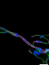
Histologically, the mammary gland is composed of a bilayered epithelium consisting of an inner layer of luminal cells (keratin 8+) and an outer layer of contractile basal cells (keratin 5+). Luminal cells are responsible for milk production during lactation, while basal cells aid milk ejection. The epithelium is surrounded by a stromal fat pad that comprises fibroblasts, nerves, vasculature, lymphatics, immune cells, adipocytes, and extracellular matrix (ECM) (Richert et al., 2000).
Over the past decade, organoids of various tissues, such as stomach, colon, lung, and pancreas, have been developed (Huch and Koo, 2015), offering many exciting possibilities for the study of tissue development and disease. The organoid system is a powerful tool that combines the advantages of a 2D culture (easy manipulation, precise control of cell composition and microenvironment, live imaging) with the opportunity to study complex cell–cell and cell–ECM interactions in a more controlled ex vivo manner (Huch and Koo, 2015; Shamir and Ewald, 2015; Koledova, 2017; Artegiani and Clevers, 2018).
Several models have been developed to study the mechanisms of mammary branching morphogenesis in primary mammary epithelium using different protocols (Ewald et al., 2008; Huebner et al., 2016; Neumann et al., 2018), cell lines (Xian et al., 2005), sorted cells (Jamieson et al., 2017; Linnemann et al., 2015), or induced pluripotent stem cells (Qu et al., 2017). However, an organoid system modeling key aspects of the late postnatal developmental stages of the mammary gland has remained challenging to establish.
Previously, there have been several attempts to model lactation in 3D culture: spheroids of a breast adenoma cell line were used to study copper secretion into milk (Freestone et al., 2014); organoids of primary epithelium were shown to produce milk following the administration of a lactogenic stimulus (Mroue et al., 2015; Jamieson et al., 2017); and co-culture of breast epithelium and pre-adipocyte cell lines was shown to initiate an involution-like process (Campbell et al., 2014). However, in-depth characterization of milk production and involution or the proper bilayered architecture of mammary epithelium remained to be carried out.
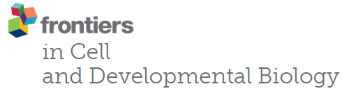
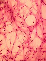
Recently, we developed a model of lactation and involution of mammary epithelium based on organoids of primary mammary gland tissue cultured in 3D Matrigel® (Sumbal et al., 2020b). Under lactogenic stimuli, primary organoids maintain long-term milk production, retain the contractile myoepithelial layer, and enter involution following hormone withdrawal. Moreover, after involution, the organoids remain hormonally sensitive and are able to enter another round of lactation (Sumbal et al., 2020b). Here, we present a methodological guideline to establish the primary mammary organoid-based ex vivo model of lactation and involution, with detailed procedures for obtaining tissue, isolating organoids, establishing and maintaining 3D culture, and preparing organoid samples for subsequent RNA or protein expression analysis or histological examination. This model can be used for studies on lactation biology, mammary stem cell plasticity, regulatory mechanisms of mammary epithelial cell differentiation and death, or other interesting biological phenomena. We believe that this model will initiate the further development of organoid technology, including creative applications in biotechnology and regenerative medicine (Sumbal et al., 2020a).
Materials and Reagents
100-mm tissue culture Petri dish (e.g., Corning, catalog number: 353003 )
0.2-μm filters and 50 ml syringes (e.g., GVS, catalog number: FJ25ASCCA002DL01 )
No. 22 disposable scalpel blades (e.g., Swann-Morton, catalog number: 0508)
50-ml tubes (e.g., Corning, catalog number: 352070 )
15-ml tubes (e.g., Corning, catalog number: 352096 )
10-ml disposable plastic pipettes (e.g., Corning, catalog number: 357551 )
25-ml disposable plastic pipettes (e.g., Corning, catalog number: 357535 )
24-well tissue culture plates (e.g., Corning, catalog number: 353047 )
30 G insulin syringes (e.g., BD Microfine, catalog number: 324826 )
Plastic histology molds (e.g., Thermo Scientific, catalog number: 1830 )
Plastic embedding cassettes (e.g., Simport, catalog number: M492-2 )
Histology tissue molds (e.g., Simport, catalog number: M474-3 )
Microscope slides for histology (e.g., Thermo Scientific, catalog number: J1800AMNZ )
Mice: virgin females, 7–10 weeks old, inbred strain C57BL/6J (e.g., The Jackson Laboratory, catalog number: 000 664 )
Ethanol (EtOH), 70%, 95%, and 100% (e.g., VWR, catalog number: 83813 )
Phosphate-buffered saline (PBS) (e.g., Sigma-Aldrich, catalog number: D1408 )
Dulbecco’s modified Eagle medium (DMEM)/F12 (e.g., Gibco, catalog number: 21331-020 )
Bovine serum albumin (BSA) (e.g., Sigma-Aldrich, catalog number: A3608 )
Fetal bovine serum (FBS) (e.g., Sigma-Aldrich, catalog number: F0804 )
Collagenase A (e.g., Roche, catalog number: 11088793001 )
Trypsin (e.g., Dutcher Dominique, catalog number: P10-022100 )
Insulin (e.g., Sigma-Aldrich, catalog number: I6634-100MG )
Gentamicin (e.g., Sigma-Aldrich, catalog number: G1397 )
Glutamine (e.g., Gibco, catalog number: 35050-061 )
DNase I (e.g., Sigma-Aldrich, catalog number: D4527-40KU )
Dispase II (e.g., Roche, catalog number: 13 75 2000 )
Growth factor-reduced Matrigel® (e.g., Corning, catalog number: 354230 )
Insulin-transferrin-selenium (ITS) (e.g., Gibco, catalog number: 41400-045 )
Penicillin/Streptomycin (e.g., Gibco, catalog number: 15140-122 )
FGF2 (e.g., Gibco, catalog number: PM60034 )
Prolactin (e.g., Sigma-Aldrich, catalog number: SRP4688 )
Hydrocortisone (e.g., Sigma-Aldrich, catalog number: S H6909 )
Oxytocin (e.g., Sigma-Aldrich, catalog number: O3251 )
RNeasy Micro Kit (e.g., Qiagen, catalog number: 74004 )
β-Mercaptoethanol (e.g., Sigma-Aldrich, catalog number: M6250 )
Phosphatase inhibitor cocktail II (e.g., Millipore, catalog number: 524625 )
RIPA buffer (e.g., Sigma-Aldrich, catalog number: R0278 )
Protease inhibitor cocktail I (e.g., Sigma-Aldrich, catalog number: 539131 )
Pierce Coomassie (Bradford) Protein Assay Kit (e.g., Thermo Scientific, catalog number: 23200 )
Paraformaldehyde (PFA), 32% (e.g., Electron Microscopy Sciences, catalog number: 15714 )
Low gelling temperature agarose (e.g., Sigma-Aldrich, catalog number: A9414 )
Xylene (e.g., Sigma-Aldrich, catalog number: 534056 )
Paraffin (e.g., Sigma-Aldrich, catalog number: 1071642504 )
Dissociation solution (see Recipes)
BSA solution (see Recipes)
Basal organoid medium (BOM) (see Recipes)
Morphogenesis medium (see Recipes)
Lactation medium (see Recipes)
4% PFA (see Recipes)
RNA lysis buffer (see Recipes)
Equipment
Surgical tools
Forceps (e.g., Phymep, catalog numbers: 11050-10 and 11051-10 )
Scissors (e.g., Phymep, catalog number: 14088-10 )
Dissection board (e.g., Thermo Scientific, catalog number: 36-119 )
P1000 pipette
Laminar flow hood
Fridge 4°C (e.g., Liebherr, catalog number: 7083 001-01 )
Freezer –80 °C (e.g., Thermo Scientific, catalog number: 88400V )
Liquid nitrogen tank (e.g., Air Liquide Espace 151, catalog number: 2433867 )
Shaking incubator at 37°C (e.g., Infors HT Multitron)
Centrifuge (e.g., Thermo Scientific, model: Sorvall ST40 )
Incubator for cell culture, 37°C, 5% CO2 (e.g., Thermo Scientific, model: HERAcell 150i )
Heating plate at 37°C (e.g., Techne DRI-Block DB-2A)
Microscope and camera (e.g., Olympus model: CKX41 )
NanoDrop™ (e.g., Implen Nanophotomoter NP80)
Sonicator (e.g., Diagenode Bioruptor Pico)
Incubator at 65°C (e.g., Memmert Incubator I)
Embedding workstation (e.g., Leica EG1150C )
Procedure
文章信息
版权信息
© 2021 The Authors; exclusive licensee Bio-protocol LLC.
如何引用
Charifou, E., Sumbal, J., Koledova, Z., Li, H. and Chiche, A. (2021). A Robust Mammary Organoid System to Model Lactation and Involution-like Processes. Bio-protocol 11(8): e3996. DOI: 10.21769/BioProtoc.3996.
分类
癌症生物学 > 通用技术 > 动物模型
细胞生物学 > 组织分析 > 组织分离
细胞生物学 > 细胞分离和培养 > 3D细胞培养
您对这篇实验方法有问题吗?
在此处发布您的问题,我们将邀请本文作者来回答。同时,我们会将您的问题发布到Bio-protocol Exchange,以便寻求社区成员的帮助。