

Imaging Microtubules in vitro at High Resolution while Preserving their Structure
保留微管结构同时在体外对其进行高分辨率成像

发布: 2021年04月05日第11卷第7期 DOI: 10.21769/BioProtoc.3968 浏览次数: 3597
评审: Thirupugal GovindarajanOneil Girish BhalalaAnonymous reviewer(s)
参见作者原研究论文
The authors used this protocol in:

Apr 2020


Abstract
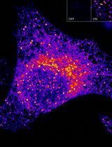
Microtubules (MT) are the most rigid component of the cytoskeleton. Nevertheless, they often appear highly curved in the cellular context and the mechanisms governing their overall shape are poorly understood. Currently, in vitro microtubule analysis relies primarily on electron microscopy for its high resolution and Total Internal Reflection Fluorescence (TIRF) microscopy for its ability to image live fluorescently-labelled microtubules and associated proteins. For three-dimensional analyses of microtubules with micrometer curvatures, we have developed an assay in which MTs are polymerized in vitro from MT seeds adhered to a glass slide in a manner similar to conventional TIRF microscopy protocols. Free fluorescent molecules are removed and the MTs are fixed by perfusion. The MTs can then be observed using a confocal microscope with an Airyscan module for higher resolution. This protocol allows the imaging of microtubules that have retained their original three-dimensional shape and is compatible with high-resolution immunofluorescence detection.
Keywords: Microtubules (微管)Background
Microtubules (MT) are polymers made by the combination of the heterodimers α- and β-tubulins and are a major component of the cell cytoskeleton. They are involved in fundamental mechanisms of cell function such as mitosis, intracellular transport, cytokinesis and maintenance of cell shape (Akhmanova and Steinmetz, 2015). Although inherently very rigid, MTs often appear curved in cells and few proteins have been described to bend microtubules (Brangwynne et al., 2006; Bechstedt et al., 2014; Leung et al., 2020; Cuveillier et al., 2020). Since the early 1970s (Weisenberg, 1972), the study of MTs in vitro has led to a better understanding of the molecular mechanism involved in the formation and dynamics of MTs. However, a detailed analysis of the shape of the microtubules remains technically difficult. Two main approaches are currently used: electron microscopy for the very detailed images obtained (up to a separation limit of a few Angstrom) (Alushin et al., 2014; Harris, 2015), and TIRF microscopy which allows the live observation of dynamic microtubules using fluorescently-labelled molecules (separation limit of about 200 nm) (Al-Bassam, 2014). However, these techniques are not suitable for the complete observation of microtubules with large three-dimensional curvatures such as the helical shape they adopt in the presence of MAP6 (Cuveillier et al., 2020). In order to obtain more clues on the structure of the MTs, we developed an assay that combines the use of fluorescent proteins and high-resolution imaging by Airyscan confocal microscopy (achievable resolution down to 120 nm in XY), while preserving the original shape of the MTs which is very sensitive to manipulation. This protocol has the advantage of avoiding unusual equipment and material. Moreover, it can be adapted to different super-resolution techniques such as expansion microscopy or stimulated-emission-depletion (STED) microscopy which would allow to increase the resolution up to 10 times (Blom and Brismar, 2014) and reach a separation limit of 50-20 nm.
Materials and Reagents
Tubulin, Atto-565 labelled tubulin, and biotinylated tubulin are prepared as already fully described in Ramirez-Rios et al. (2017). Store up to 1 year in liquid nitrogen
Cover glasses 26 × 76 mm #1 (VWR, catalog number: 630-2910 )
Double-face precut tape 70 µm thick, 3 mm wide (LIMA Company, catalog number: 0000P70PC3003 )
Siligum wax plate (VWR, MODU140013 )
1.5 ml Eppendorf tubes (Fisher Scientific, catalog number: 11558232 )
0.5 ml Eppendorf tubes (Fisher Scientific, catalog number: 10318661 )
Petri dishes (Greiner Bio-One, catalog number: 663102 )
Polycarbonate centrifuge tubes (Beckman, catalog number: 343775 )
0.22 µm filters (Merck Millipore, catalog number: SLGP033RS )
Silane-PEG (MW 30k) (Creative PEG-Works, catalog number: PSB-2014 )
Silane-PEG-biotin (MW 3,400) (LaysanBio, catalog number: BIOTIN-PEF-SIL , MW 3,400)
NeutrAvidin (ThermoScientific, catalog number: 31000 )
PLL20K-G35-PEG2K (PLL-g-PEG) (Jenkem, catalog number: 13022755 )
Pluronic F-127 (Sigma-Aldrich, catalog number: P2443 )
Bovine Serum Albumin (BSA) (Sigma-Aldrich, catalog number: A7030 )
Acetone 100% (VWR, catalog number: 20066.321 )
Ethanol 96% (VWR, catalog number: 20823362 )
Hellmanex III (Sigma-Aldrich, catalog number: Z805939-1EA )
Phosphate buffer saline (PBS) (Sigma-Aldrich, catalog number: P4417 )
1,4-Piperazinediethanesulfonic acid (PIPES) (Sigma-Aldrich, catalog number: P6757 )
Potassium hydroxide (KOH) (Sigma-Aldrich, catalog number: 484016 )
Potassium chloride (KCl) (Sigma-Aldrich, catalog number: P9541 )
Ethylene glycol-bis(2-aminoethylether)-N,N,N′,N′-tetraacetic acid (EGTA) (Sigma-Aldrich, catalog number: E3889 )
Magnesium chloride (MgCl2) (Sigma-Aldrich, catalog number: 2670 )
Sodium hydroxide (NaOH) (Carlo Erba, catalog number: 480507 )
DL-Dithiothreitol (DTT) (Sigma-Aldrich, catalog number: D0632 )
Guanosine 5′-triphosphate (GTP) (Sigma-Aldrich, catalog number: G8877 )
Methyl cellulose 1,500 cP (Sigma-Aldrich, catalog number: M0387 )
GMPCPP (Euromedex, JE-NU-405S )
HEPES (Sigma-Aldrich, catalog number: H3375 )
Glucose (Sigma-Aldrich, catalog number: G8270 )
Glucose oxidase (Sigma-Aldrich, catalog number: G6766-10KU )
Catalase (Sigma-Aldrich, catalog number: C9322-1G )
Diamond pencil (Agar Scientific, catalog number: AGT5347 )
Glutaraldehyde solution 25% (Sigma-Aldrich, catalog number: G5882 )
Clean nitrogen air flow (Air Liquide)
Liquid nitrogen (Air Liquide)
5× BRB80 (see Recipes)
BSA 10% (see Recipes)
Neutravidine (see Recipes)
DTT, 200 mM (see Recipes)
KCl, 500 mM (see Recipes)
PLL-PEG (see Recipes)
Silane-PEG or Silane-PEG-biotin (see Recipes)
GTP, 20 mM (see Recipes)
Glucose, 450 mg/ml (see Recipes)
Deoxymix (catalase and glucose oxydase) (see Recipes)
NaOH, 1 M (see Recipes)
BSA 1%/BRB80 (see Recipes)
Methyl cellulose 1,500 cP (see Recipes)
HEPES, 10 mM (see Recipes)
Pluronic F27, 10% (see Recipes)
PBS (see Recipes)
Neutravidin stock solution (see Recipes)
Red tubulin mix (see Recipes)
Imaging buffer (see Recipes)
Equipment
Inverted Eclipse Ti microscope (Nikon) with PSF focus
Apochromat 100×/1.49 N.A. oil immersion objective (Nikon) with heated objective (Okolab)
Temperature chamber (Technico Plast)
Ilas2 TIRF system (Roper Scientific)
Cooled Charged-coupled device camera ( Evolve 512 , Photometrics)
Temperature Stage Controller (Linkam Scientific)
LSM 710 confocal (Zeiss) Airyscan
Plan Apochromat 100×/1.4 N.A. oil immersion objective (Zeiss)
Ultracentrifuge (Beckman Coulter Optima, Model Max-XP, catalog number: 393315 )
Rotor (Beckman, model: TLA100-1, catalog number: 343837)
Plasma cleaner FEMTO Diener Electric (Germany) coupled to a vacuum pump Trivac D2.5E Oerliken (Germany)
Sonicator ( Elmasonic S30 , Elma, Germany)
Glass staining dishes and tray (VWR, catalog number: MARI4200004 )
Plastic box for glass microscopy (any brand)
Diamond Pencil (Oxford Instruments, catalog number: T5347 )
Tweezers (Dutscher, catalog numbers: 076100 and 005093 )
Gloves powder-free (any brand)
Water-bath (any brand)
Software
MetaMorph 7.8.5 software (Molecular Devices, https://www.moleculardevices.com)
Zen Black 2.1 (Zeiss)
Procedure
文章信息
版权信息
© 2021 The Authors; exclusive licensee Bio-protocol LLC.
如何引用
Readers should cite both the Bio-protocol article and the original research article where this protocol was used:
- Cuveillier, C., Saoudi, Y., Arnal, I. and Delphin, C. (2021). Imaging Microtubules in vitro at High Resolution while Preserving their Structure. Bio-protocol 11(7): e3968. DOI: 10.21769/BioProtoc.3968.
- Cuveillier, C., Delaroche, J., Seggio, M., Gory-Fauré, S., Bosc, C., Denarier, E., Bacia, M., Schoehn, G., Mohrbach, H., Kulić, I., Andrieux, A., Arnal, I. and Delphin, C. (2020). MAP6 is an intraluminal protein that induces neuronal microtubules to coil. Sci Adv 6(14): eaaz4344.
分类
神经科学 > 细胞机理
生物化学 > 蛋白质 > 成像
生物化学 > 蛋白质 > 荧光
您对这篇实验方法有问题吗?
在此处发布您的问题,我们将邀请本文作者来回答。同时,我们会将您的问题发布到Bio-protocol Exchange,以便寻求社区成员的帮助。