

Intestinal Co-culture System to Study TGR5 Agonism and Gut Restriction
利用肠道共培养系统研究TGR5激动剂和肠道限制性
发布: 2021年03月20日第11卷第6期 DOI: 10.21769/BioProtoc.3948 浏览次数: 6653
评审: Pierre-Damien DenechaudAnonymous reviewer(s)
参见作者原研究论文
The authors used this protocol in:

Jan 2021


Abstract
The activation of the Takeda G-protein receptor 5 (TGR5, also known as the G protein-coupled bile acid receptor 1, GPBAR1) in enteroendocrine L-cells results in secretion of the anti-diabetic hormone Glucagon-Like Peptide 1 (GLP-1) into systemic circulation. Consequently, recent research has focused on identification and development of TGR5 agonists as type 2 diabetes therapeutics. However, the clinical application of TGR5 agonists has been hampered by side effects of these compounds that primarily result from their absorption into circulation. Here we describe an in vitro screening protocol to evaluate the TGR5 agonism, GLP-1 secretion, and gut-restricted properties of small molecules. The protocol involves differentiating gut epithelial and endocrine cells together in transwells to assess both the pharmacodynamics of TGR5 agonists and the toxicity of compounds to the intestinal monolayer. As a proof of concept, we demonstrate the use of the protocol in evaluating properties of naturally occurring bile acid metabolites that are potent TGR5 agonists. This protocol is adapted from Chaudhari et al. (2021).
Keywords: Enteroendocrine cells (肠内分泌细胞)Background
The intestinal wall of the GI tract is comprised of several different types of cells, each with specific and, sometimes exclusive functions (Allaire et al., 2018). In vitro testing of small molecule therapeutics or drug candidates has traditionally relied on mono-enteric cell lines. These in vitro systems do not determine the absorptive properties of the small molecule being tested, nor do they predict the behavior of these compounds in the gut. To some extent, enteroids recapitulate the complexity and diversity of the intestine, but they are an expensive system and do not always provide reproducible results due to variability in the reagents required (Blutt et al., 2018). There is an unmet need for simple experimental systems that mimic the gut epithelial layer to aid in the development of new chemical probes and drugs. Here, we focus on developing cell-based protocols to assay one intestinal signaling system, the small molecule-based activation of the G protein-coupled receptor TGR5 on intestinal enteroendocrine cells.
The study of small molecule agonists of TGR5 has gained significant importance in recent years due to the potential therapeutic benefits of these compounds, primarily in diabetes control (Duboc et al., 2014). TGR5 is expressed on enteroendocrine L-cells in the lower gastrointestinal (GI) tract, and when activated, results in secretion of the anti-diabetic hormone GLP-1 into the bloodstream (Eissele et al., 1992; Harach et al., 2012). Activation of this gut-initiated TGR5-GLP1 cascade shows promise as a potential target for type 2 diabetes therapeutics (Hodge and Nunez, 2016). However, activation of TGR5 outside of the gut can cause unwanted side effects (Cao et al., 2016; Hodge and Nunez, 2016). Therefore, a unified in vitro screening method that can be used to evaluate the effects of small molecules on TGR5 activation and GLP-1 secretion, as well as the interaction of these compounds with the gut epithelial barrier, will aid in the development of successful new TGR5-targeted therapies.
Here we describe a simple in vitro protocol from Chaudhari et al. (2021) to co-differentiate human intestinal epithelial cells with human enteroendocrine cells into a polarized monolayer in transwells. We then describe how to utilize the resulting epithelial layer to test the TGR5 agonism and gut restricted-properties of small molecules. Our system incorporates GLP-1-secreting NCI-H716 L-cells, which have been the predominant cell line used to test TGR5 activation and GLP-1 secretion (Kim et al., 2017; Sato et al., 2008), into an epithelial monolayer formed by surrounding human intestinal colon carcinoma Caco-2 epithelial cells. This monolayer forms a physical and biochemical barrier to small molecules.
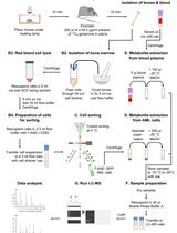
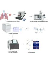
Through electron microscopy imaging, we show that the enteroendocrine cells intercalate with the colon epithelial cells and are polarized, with distinct apical and basolateral faces. Mass spectrometry-based quantification of the passage of small molecules from the apical to the basolateral chamber through the differentiated monolayer allows for evaluation of the gut-restricted properties of the compounds (Chaudhari et al., 2021). Using this system, we can also perform RNA interference knockdowns, harvest cells to extract RNA and quantify TGR5 expression, and measure GLP-1 secretion. Importantly, because small molecules can be added to either face of the differentiated monolayer, information about whether compounds can induce TGR5 activation and expression and GLP-1 secretion from the apical face, basolateral face, or both faces, can be determined. Lastly, we describe how this system can be used to study toxic effects of small molecules on intestinal cells as well as the effects of these compounds on epithelial barrier integrity. This in vitro co-culture transwell system will enable the identification of small molecules that are both TGR5 agonists and possess limited off-targeted effects and will thus facilitate the development of novel type 2 diabetes therapies.
Materials and Reagents
TC treated flasks (Genesee, catalog number: 25-210 )
Olympus vacuum filter systems, 500 ml (Genesee, catalog number: 25-227 )
Falcon 15 ml conical centrifuge tubes (Fisher Scientific, catalog number: 14-959-53A )
Falcon 50 ml conical centrifuge tubes (Fisher Scientific, catalog number: 14-432-22 )
Corning Transwell polycarbonate membrane cell culture inserts (Millipore Sigma, catalog number: CLS3401-48EA )
RNase-free microfuge tubes (1.5 ml) (Fisher Scientific, catalog number: AM12400 )
Precellys lysing tubes VK05, 2 ml (Bertin Instruments, catalog number: P000913-LYSK0-A )
Sep-Pak C-18 40 mg 96-well pate (Waters, catalog number: 186003966 )
Fisherbrand 96-Well DeepWell polypropylene microplates (Fisher Scientific, catalog number: 12-566-120 )
Corning 96-well solid black polystyrene microplates (Fisher Scientific, catalog number: 07-200-590 )
Human colorectal adenocarcinoma cell line Caco-2 (ATCC, catalog number: HTB-37 )
Human colorectal adenocarcinoma cell line NCI-H716 (ATCC, catalog number: CCL-251 )
DMEM, high glucose, with L-glutamine (Genesee, catalog number: 25-500 )
RPMI 1640, with L-glutamine and phenol red (Genesee, catalog number: 25-506 )
GenClone fetal bovine serum (Genesee, catalog number: 25-514 )
Penicillin-streptomycin 100× solution (Genesee, catalog number: 25-512 )
Trypsin-EDTA, 0.25% 1×, phenol red (Genesee, catalog number: 25-510 )
Gibco Trypan blue solution, 0.4% (Thermo Fisher Scientific, catalog number: 15250061 )
Fluorescein isothiocyanate-dextran 4 kDa (Millipore Sigma, catalog number: 46944-100MG-F )
Opti-MEM reduced serum media, GlutaMAX Supplement (Thermo Fisher Scientific, catalog number: 51985091 )
Lipofectamine 3000 transfection reagent (Thermo Fisher Scientific, catalog number: L3000001 )
PBS 1× (Genesee, catalog number: 25-507 )
Trifluoroacetic acid (TFA) (Millipore Sigma, catalog number: T6508 )
Hydrochloric acid (HCl) (Millipore Sigma, catalog number: H1758 )
Formic acid (Millipore Sigma, catalog number: F0507 )
NaCl (Millipore Sigma, catalog number: S9888 )
Isopropyl alcohol (Millipore Sigma, catalog number: 563935 )
GLP-1 EIA Kit (Millipore Sigma, catalog number: RAB0201-1KT )
TRIzol reagent (Thermo Fisher Scientific catalog number: 155626 )
Promega CellTiter-Glo luminescent cell viability assay kit (Promega, catalog number: PR-G7570 )
MTT Assay for cell viability and proliferation (Millipore Sigma, catalog number: 11465007001 )
Complete media (see Recipes)
Transfection media (see Recipes)
Low-serum media for GLP-1 secretion (see Recipes)
Cell lysis buffer (see Recipes)
Equipment
Hemocytometer for cell counting (Weber Scientific, catalog number: 3048-12)
-80 °C freezer
Biosafety cabinet
Water bath (37 °C)
Tissue culture incubator (37 °C, 5% CO2)
Inverted microscope (Amscope)
Bench-top centrifuge
Fluorescence and absorbance plate reader (FlexStation 3, Molecular Devices)
MagNA Lyser (Roche Diagnostics)
SpeedVac vacuum concentrators (Thermo Fisher Scientific)
Ultra-high performance liquid chromatography-mass spectrometry (UPLC-MS) with a 6120 Quadrupole LC/MS spectrometer (Agilent Technologies)
qPCR instrument (QuantStudio 7 Pro, Thermo Fisher Scientific)
Software
SoftMax Pro 7 (Molecular Devices, http://www.moleculardevices.com)
OpenLab Chromatography Data System (Agilent Technologies)
Applied Biosystems qPCR analysis software (QuantStudio 7 Pro, Thermo Fisher Scientific)
Microsoft Excel and GraphPad Prism 7 (https://www.graphpad.com/scientific-software/prism/) for data plotting and analyses
Procedure
文章信息
版权信息
© 2021 The Authors; exclusive licensee Bio-protocol LLC.
如何引用
Chaudhari, S. N. and Devlin, A. S. (2021). Intestinal Co-culture System to Study TGR5 Agonism and Gut Restriction. Bio-protocol 11(6): e3948. DOI: 10.21769/BioProtoc.3948.
分类
癌症生物学 > 癌症生物化学 > 癌代谢
细胞生物学 > 细胞新陈代谢 > 糖类
您对这篇实验方法有问题吗?
在此处发布您的问题,我们将邀请本文作者来回答。同时,我们会将您的问题发布到Bio-protocol Exchange,以便寻求社区成员的帮助。