

Generation of the Compression-induced Dedifferentiated Adipocytes (CiDAs) Using Hypertonic Medium
使用高渗培养基压缩诱导的去分化脂肪细胞(CiDAs)的产生
发布: 2021年02月20日第11卷第4期 DOI: 10.21769/BioProtoc.3920 浏览次数: 8772
评审: Shengwen Calvin LiNandini MondalAnonymous reviewer(s)
参见作者原研究论文
The authors used this protocol in:
Jan 2020
Advertisement


Abstract
Current methods to obtain mesenchymal stem cells (MSCs) involve sampling, culturing, and expanding of primary MSCs from adipose, bone marrow, and umbilical cord tissues. However, the drawbacks are the limited numbers of total cells in MSC pools, and their decaying stemness during in vitro expansion. As an alternative resource, recent ceiling culture methods allow the generation of dedifferentiated fat cells (DFATs) from mature adipocytes. Nevertheless, this process of spontaneous dedifferentiation of mature adipocytes is laborious and time-consuming. This paper describes a modified protocol for in vitro dedifferentiation of adipocytes by employing an additional physical stimulation, which takes advantage of augmenting the stemness-related Wnt/β-catenin signaling. Specifically, this protocol utilizes a polyethylene glycol (PEG)-containing hypertonic medium to introduce extracellular physical stimulation to obtain higher efficiency and introduce a simpler procedure for adipocyte dedifferentiation.
Keywords: Mesenchymal stem cells (间充质干细胞)Background
Adipose tissue currently is one of the most appealing sources of mesenchymal stem cells (MSCs), due to its large abundance and relatively less-invasive harvest methods (Shen et al., 2011; González-Cruz et al., 2012; Konno et al., 2013). Adipose-derived MSCs, that isolated from the stromal-vascular fraction of subcutaneous adipose tissue, have been demonstrated to display multilineage potentials both in vitro and in vivo (Anghileri et al., 2008; González et al., 2009; Gonzalez-Rey et al., 2010; Jumabay et al., 2010; Mao et al., 2017 and 2019; Darnell et al., 2018). To isolate adipose-derived MSCs, the widely-used method is to dissect the stromal-vascular fraction from the adipose tissue, and then sort the MSCs by either fluorescence-activated cell sorting (FACS) or culture selection (Aronowitz et al., 2015; Raposio et al., 2017; Gentile et al., 2019). However, heterogeneous groups of cells are contained in a stromal-vascular fraction of adipose tissue, and limited cell markers are available for MSCs selection; these make it difficult to purify adipose-derived MSCs (Gimble et al., 2011; González-Cruz et al., 2012; Konno et al., 2013).
Alternatively, the adipocytes, rather than the other types of cells in adipose tissue, can spontaneously dedifferentiate into multipotent mesenchymal cells named the dedifferentiated fat (DFAT) cells during in vitro culturing (Sugihara et al., 1986; Shen et al., 2011; Taniguchi et al., 2016). Because of the multipotency of the DFAT cells and the large abundance of the mature adipocytes, the DFAT cells have been regarded as an ideal source for human postnatal mesenchymal multipotent stem cells (Matsumoto et al., 2008; Shen et al., 2011; Côté et al., 2019). However, the current ceiling culturing for adipocyte dedifferentiation requires a long duration (typically 4 weeks) to enable the adipocytes to spontaneously lose all obvious lipid droplets (Lessard et al., 2015; Taniguchi et al., 2016). Thus, further increasing the efficiency of adipocyte dedifferentiation and shortening its processing time is attractive for its wider applications.
Adipocytes and adipose progenitor cells are also important components in tumor microenvironments (Chandler et al., 2012; Seo et al., 2015; Ling et al., 2020). Recent studies revealed that the dedifferentiation of adipocytes occurred during tumor development, which might be attributed to the activated Wnt signaling (Gustafson and Smith, 2010; Bochet et al., 2013) and Notch signaling (Bi et al., 2016). Recent studies also revealed that the dedifferentiation of adipocytes could occur in vivo in mice models (Bochet et al., 2013; Liao et al., 2015; Wang et al., 2018). Tumor progression also largely alters the local physical microenvironments, including elevated osmotic pressure, increased compressive force, and matrix stiffening (Nia et al., 2020). These physical cues largely influence the cell fates of both adipose stromal cells and cancer cells (Guo et al., 2017; Li et al., 2019 and 2020a; Han et al., 2020). Indeed, our recent study reported that the generation of osmotic stress in vitro to mimic the elevated osmolarity in in vivo tumors could also induce the dedifferentiation of adipocytes (Li et al., 2020b). Consistently, another study also reported that a tough implant in vivo drove the dedifferentiation of the local surrounding adipocytes (Ma et al., 2019). Thus, these studies inspired us to develop an alternative protocol to generate multipotent mesenchymal cells by mechanically dedifferentiating adipocytes.
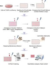
The protocol described here includes the experimental set-ups to induce and verify the reprogramming of adipocytes into multipotent mesenchymal cells using our hypertonic dedifferentiation medium. We also include the procedures to generate adipocytes from preadipocytes or mesenchymal stem cells, and the differentiation assays to confirm the multilineage potentials of the CiDAs.
Materials and Reagents
Reagents
Minimum Essential Medium Eagle Alpha Modification media (Sigma-Aldrich, catalog number: M8042 )
Fetal bovine serum (Gibco, catalog number: 10-082-147 )
Penicillin/streptomycin (Gibco, catalog number: 15140148 )
Polyethylene glycol 300 (Sigma-Aldrich, catalog number: 8.07484 )
KnockOut Serum Replacement (Gibco, catalog number: 10828-028 )
Preadipocyte Growth Medium-2 (Lonza, catalog number: PT-8202 )
SingleQuots (Lonza, catalog number: PT-9502 )
Paraformaldehyde (VWR, catalog number: IC0219998380 )
PBS (Sigma-Aldrich, catalog number: P5119 )
Triton-X-100 (Sigma-Aldric, catalog number: X100 )
Oil Red O (Sigma-Aldrich, catalog number: O0625 )
DMEM (Sigma-Aldrich, Brand, catalog number: D5546 )
Horse serum (Gibco, catalog number: 26050070 )
Dexamethasone (Sigma-Aldrich, catalog number: D4902 )
Hydrocortisone (Sigma-Aldrich, catalog number: H0888 )
Hydrogen peroxide (Sigma-Aldrich, catalog number: H1009 )
Anti-MyoD1 (Abcam, catalog number: ab16148 )
Donkey anti-Rabbit Alexa 488 (Invitrogen, catalog number: R37118 )
β-glycerophosphate (Sigma-Aldrich, catalog number: G9422 )
L-ascorbic acid (Sigma-Aldrich, Brand, catalog number: A4403 )
ELF-97 (Invitrogen, catalog number: E6588 )
TGF-β (R&D Systems, catalog number: 240-B )
Anti-aSMA (Abcam, catalog number: ab5694 )
DAPI (Thermo Scientific, Brand, catalog number: 62248 )
Trypsin (2.5%) (Thermo Fisher Scientific, GibcoTM, catalog number: 15090046 )
Cell culture plasticware
T75 and/or T25 flasks (Corning, catalog numbers: 430641U for T75 and 3056 for T25)
Centrifuge tubes (15 ml; 50 ml, Corning, catalog numbers: 430790 ; 430828 )
Cryovials (STARLAB, catalog number: E3110-6122 )
Pipette tips (TipOne, STARLAB, catalog numbers: S1111-3700 ; S1111-1706 ; S1111-6701 )
35-mm cell culture dish (Thermo Fisher Scientific, catalog number: 153066 )
6-well plates (Corning, Falcon®, catalog number: 353934 )
100 mm cell culture dish (Thermo Fisher Scientific, Thermo ScientificTM, catalog number: 150464 )
Equipment
Centrifuge (Eppendorf, model: 5810 )
Bright-LineTM Hemacytometer (Sigma-Aldric, catalog number: Z359629 )
Water bath (Thermo Scientific, catalog number: TSCIR19 )
Humidified incubator at 37 °C, 5% CO2 (Thermo Fisher Scientific, Heraeus, model: HeracellTM 150)
Xenon Arc Lamp ( ZEISS )
Hamamatsu Orca Flash 4.0 V3 ( Scientifica )
Aspirator (Dry vacuum pump/compressor, Welch Vacuum - Gardner Denver, model: 2511 )
Software
ImageJ (https://imagej.nih.gov/ij/)
LAS X ( Leica Microsystems, Mannheim, Germany)
HCImage (http://www.hamamatsu.com/)
Procedure
文章信息
版权信息
© 2021 The Authors; exclusive licensee Bio-protocol LLC.
如何引用
Readers should cite both the Bio-protocol article and the original research article where this protocol was used:
- Li, Y., Mao, A. S., Seo, B. R., Zhao, X., Gupta, S. K., Chen, M., Han, Y. L., Shih, T., Mooney, D. J. and Guo, M. (2021). Generation of the Compression-induced Dedifferentiated Adipocytes (CiDAs) Using Hypertonic Medium. Bio-protocol 11(4): e3920. DOI: 10.21769/BioProtoc.3920.
- Li, Y., Mao, A. S., Seo, B. R., Zhao, X., Gupta, S. K., Chen, M., Han, Y. L., Shih, T. Y., Mooney, D. J. and Guo, M. (2020). Compression-induced dedifferentiation of adipocytes promotes tumor progression. Sci Adv 6(4): eaax5611.
分类
干细胞 > 成体干细胞 > 脂肪干细胞
癌症生物学 > 通用技术 > 细胞生物学试验
您对这篇实验方法有问题吗?
在此处发布您的问题,我们将邀请本文作者来回答。同时,我们会将您的问题发布到Bio-protocol Exchange,以便寻求社区成员的帮助。