

Relative Quantification of NaV1.1 Protein in Mouse Brains Using a Meso Scale Discovery-Electrochemiluminescence (MSD-ECL) Method
利用中尺度发现电化学发光(MSD-ECL)方法对小鼠脑中NaV1.1蛋白进行相对定量
发布: 2021年02月05日第11卷第3期 DOI: 10.21769/BioProtoc.3910 浏览次数: 4668
评审: Dhruv Rajanikant PatelAnca Flavia SavulescuAnonymous reviewer(s)

参见作者原研究论文
The authors used this protocol in:
Aug 2020
Advertisement


Abstract
Densitometric analysis is often used to quantify NaV1.1 protein on immunoblots, although the sensitivity and dilution linearity of the method are usually poor. Here we present a protocol for quantification of NaV1.1 in mouse brain tissues using a Meso Scale Discovery-Electrochemiluminescence (MSD-ECL) method. MSD-ECL is based on ELISA (enzyme-linked immunosorbent assay) and uses electrochemiluminescence to produce measurable signals. Two different antibodies are used in this assay to capture and detect NaV1.1 respectively in brain tissue lysate. The specificity of the antibodies is confirmed by Scn1a gene knock-out tissue. The calibration curve standards used in this assay were generated with mouse liver lysate spiked with mouse brain lysate, instead of using a recombinant protein. We showed that this method was qualified and used for quantification of NaV1.1 in mouse brain tissues with specificity, accuracy and precision.
Keywords: NaV1.1 (NaV1.1)Background
NaV1.1, also known as the alpha subunit of voltage gated sodium channel, type I, is a transmembrane protein encoded by the Scn1a gene (Meisler et al., 2010). Decreased expression of functional NaV1.1 leads to Dravet syndrome (DS), a severe early-onset epileptic encephalopathy (Dravet et al., 2005). Expression of NaV1.1 in biological samples has been used as a non-clinical pharmacological biomarker for DS and can be measured using densitometric analysis of immunoblots. Densitometry methods are often not as accurate and sensitive as standard immunoassays. In addition, some NaV1.1 antibodies may cross react with other voltage gated sodium channels (VGSCs), including NaV1.2, NaV1.3 and NaV1.8, due to homology of the protein sequences. We developed a specific and sensitive method to quantify NaV1.1 protein in mouse brain tissue using a Meso Scale Discovery-Electrochemiluminescence (MSD-ECL) method. MSD-ECL method is ELISA (enzyme-linked immunosorbent assay) based and uses electrochemiluminescence to produce measurable signals based on labeled antibody binding to the target (Kuhle et al., 2016). Scheme of antibody binding in MSD-ECL assay described in this protocol is shown in Figure 1. The MSD-ECL method has advantages of low background and high sensitivity. The specificity of this assay was confirmed by using capture and detection antibodies that were validated using Scn1a knock-out (Scn1a-/-) mouse brain tissue (Figure 2). Since NaV1.1 is a transmembrane protein, reproducible production of a high-quality reference standard in cells could be challenging. Therefore, in this method, reference standards composed of mouse liver lysate spiked with mouse brain lysate was utilized. Mouse liver lysate was selected as a diluent to maintain equivalent protein load in each standard since it did not show detectable levels of NaV1.1 protein expression (Figure 3). This “fit for purpose” MSD-ECL method was qualified and used for quantification of NaV1.1 in mouse brain tissues with specificity, accuracy and precision. This method is limited, however, to have a relative narrow dynamic range of detection: The upper limit of quantification (ULOQ) is set to 100% of the NaV1.1 expression in adult mouse brain. Further dilution of some sample lysate may be necessary if the NaV1.1 expression in those samples exceed the ULOQ in the initial run.
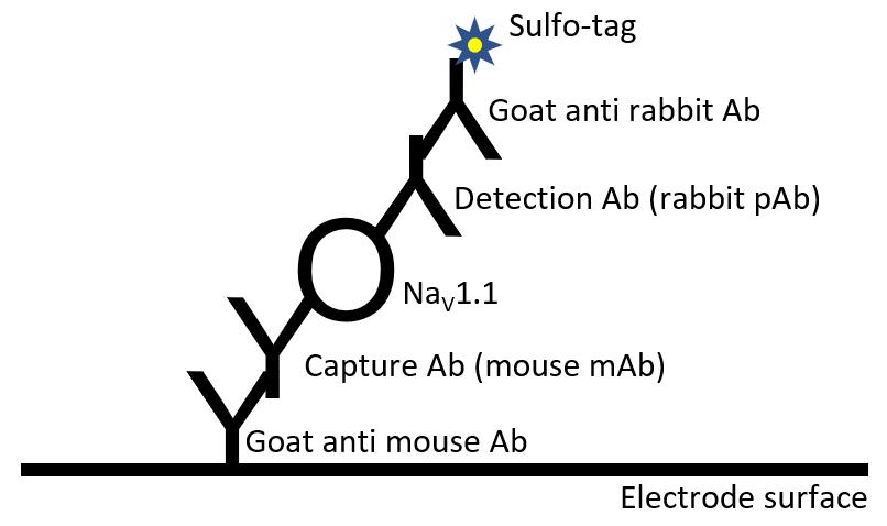
Figure 1. Scheme of antibody binding in MSD-ECL assay described in this protocol. The electrode surface of each well is pre-coated with goat anti-mouse antibody (Ab). A mouse monoclonal antibody (mAb) is used as capture Ab for NaV1.1. A rabbit polyclonal antibody (pAb) is used as detection Ab. The capture and detection antibodies bind to different epitopes within NaV1.1. A Sulfo-tag labeled goat anti-rabbit Ab produces electrochemiluminescence signal.
Materials and Reagents
2-ml Eppendorf microcentrifuge tubes, LoBind for protein (Fisher Scientific, catalog number: 13-698-795 )
1.5-ml Eppendorf microcentrifuge tubes, LoBind for protein (Fisher Scientific, catalog number: 13-698-794 )
0.2 µm nitrocellulose membrane (GE Healthcare Life Sciences, catalog number: 10600004 )
MULTI-ARRAY 96 Small Spot GAM Plate (Meso Scale Discovery, catalog number: L45MA-2 )
Scn1atm1Kea 129S6.Scn1a+/− mice (The Jackson Laboratory, catalog number: 37107-Jax )
Liquid nitrogen
PageRuller Plus prestained protein ladder (Thermo Fisher Scientific, catalog number: 26620 )
Halt protease inhibitor cocktail (100×) (Thermo Fisher Scientific, catalog number: 78429 )
Capture antibody, mouse anti NaV1.1 monoclonal antibody (NeuroMab, catalog number: 75-023 )
Detection antibody, rabbit anti NaV1.1 polyclonal antibody (Alomone, catalog number: ASC-001 )
Anti-Rabbit antibody, Goat, Sulfo-Tag labeled (Meso Scale Discovery, catalog number: R32AB-1 )
Anti-vinculin antibody (E1E9V), rabbit monoclonal antibody (Cell Signaling Technology, catalog number: 13901 )
Phosphate-buffered saline (PBS), pH 7.4 (Thermo Fisher Scientific, catalog number: 10010023 )
TGX acrylamide gradient gel (Bio-Rad, catalog number: 5671124 )
ECL Prime blocking reagent (GE Healthcare Life Sciences, catalog number: RPN418V )
ECL Plus Western Blotting Substrate (Thermo Fisher Scientific, catalog number: 32132 )
MSD Blocker B, 2 grams (Meso Scale Discovery, catalog number: R93BB-2 )
MSD read buffer T (4×) (Meso Scale Discovery, catalog number: R92TC-2 )
TBS, 10× (Bio-Rad, catalog number: 170-6435 )
10% Tween-20 (Bio-Rad, catalog number: 1610781 )
TX-100 (Sigma-Aldrich, catalog number: T8787-100ML )
Nonidet P-40, IGEPAL CA-630 (Sigma-Aldrich, catalog number: 56741-250ML-F )
Sodium deoxycholate (Sigma-Aldrich, catalog number: D6750-25G )
0.5 M EDTA pH 8.0 (Ambion, catalog number: AM9260G )
BCA protein assay kit (Pierce, catalog number: 23227 )
MSD lysis buffer (see Recipes and Note 1)
Equipment
Meso Quickplex SQ 120 (Meso Scale Discovery, model: SQ120)
Microplate shaker (Corning, model: S2020-P4-COR )
Analytical balance (Mettler Toledo, model: AB54-S )
Cryo-cup grinder (Biospec, model: 206 )
Teflon coated pestle and mortar tissue grinder Size A. Chamber volume 10 ml (Thomas Scientific, model: 3431D76 )
Teflon coated pestle and mortar tissue grinder Size B. Chamber volume 30 ml (Thomas Scientific, model: 3431D88 )
5-speed drill press (Wen, catalog number: 4208 )
Refrigerated Centrifuge (Eppendorf, model: 5430R )
Dry Blotting System (Thermo Fisher Scientific, model: iBlot 2 )
Laser scanner (GE Healthcare Life Sciences, model: Typhoon FLA 9500 )
Software
MSD Discovery Workbench 4.0 (Meso Scale Diagnostics, LLC., www.mesoscale.com)
GraphPad Prism 8.0 (GraphPad Software, www.graphpad.com)
Procedure
文章信息
版权信息
© 2021 The Authors; exclusive licensee Bio-protocol LLC.
如何引用
Readers should cite both the Bio-protocol article and the original research article where this protocol was used:
- Han, Z., Christiansen, A., Meena, M. and Liau, G. (2021). Relative Quantification of NaV1.1 Protein in Mouse Brains Using a Meso Scale Discovery-Electrochemiluminescence (MSD-ECL) Method. Bio-protocol 11(3): e3910. DOI: 10.21769/BioProtoc.3910.
- Han, Z., Chen, C., Christiansen, A., Ji, S., Lin, Q., Anumonwo, C., Liu, C., Leiser, S. C., Meena, Aznarez, I., Liau, G. and Isom, L. L. (2020). Antisense oligonucleotides increase Scn1a expression and reduce seizures and SUDEP incidence in a mouse model of Dravet syndrome. Sci Transl Med 12(558): eaaz6100.
分类
神经科学 > 神经系统疾病 > 癫痫
生物化学 > 蛋白质 > 定量
您对这篇实验方法有问题吗?
在此处发布您的问题,我们将邀请本文作者来回答。同时,我们会将您的问题发布到Bio-protocol Exchange,以便寻求社区成员的帮助。