

Histochemical Staining of Suberin in Plant Roots
植物根部软木脂的组织化学染色
发布: 2021年02月05日第11卷第3期 DOI: 10.21769/BioProtoc.3904 浏览次数: 7862
评审: Samik BhattacharyaGanesh Chandrakant NikaljeMather A Khan
参见作者原研究论文
The authors used this protocol in:
Oct 2019
Advertisement


Abstract
Histological stains are useful tools for characterizing cell shape, arrangement and the material they are made from. Stains can be used individually or simultaneously to mark different cell structures or polymers within the same cells, and to visualize them in different colors. Histological stains can be combined with genetically-encoded fluorescent proteins, which are useful for understanding of plant development. To visualize suberin lamellae by fluorescent microscopy, we improved a histological staining procedure with the dyes Fluorol Yellow 088 and aniline blue. In the complex plant organs such as roots, suberin lamellae are deposited deep within the root on the endodermal cell wall. Our procedure yields reliable and detailed images that can be used to determine the suberin pattern in root cells. The main advantage of this protocol is its efficiency, the detailed visualization of suberin localization it generates in the root, and the possibility of returning to the confocal images to analyze and re-evaluate data if necessary.
Keywords: Suberin (软木脂)Background
Suberin is a complex polyester that, together with cutin and lignin, forms a physical barrier in land plants. In Arabidopsis (Arabidopsis thaliana), suberin primarily accumulates in the cell walls of epidermal leaf cells, in the seed coat, in the periderm and in the cell wall of root endodermal cells. In the root, suberin deposition is dynamic process (Barberon et al., 2016; Andersen et al., 2018; Ursache et al., 2020) that is modulated by various nutritional stresses, drought stress, and phytohormone signaling pathways (Baxter et al., 2009; Kosma et al., 2014; Barberon et al., 2016). Suberin in root endodermal cells acts as a barrier that controls the uptake of water and nutrients (Baxter et al., 2009; Krishnamurthy et al., 2009 and 2011; Wang et al., 2019), but also provides a protective layer against various pathogens (Thomas et al., 2007; Ranathunge et al., 2008; Holbein et al., 2019; Zhou et al., 2020; Emonet et al., 2020). Moreover, suberin is also involved in periderm formation, the tissue that envelops secondary stems as part of the bark. Given its position deep within the root, it is technically challenging to study suberin. Developing a reliable tool to study the multifunctional activities of suberin and its precise localization in cells would contribute to new discoveries in many areas of plant research. A protocol for suberin staining was described previously by Lux et al. (2005) and subsequently modified over the years (Naseer et al., 2012; Fujita et al., 2020; Ursache et al., 2020). Here, we provide a standardized protocol to stain suberin and to acquire data from 10-15 roots at a single tile-scan image by confocal microscopy in a short time. This protocol will enhance the efficiency of data generation with possibility to re-analyze data if necessary.
Materials and Reagents
Square dishes 120 × 120 × 17 mm (Greiner Bio-One GmbH, catalog number: 688102 )
Flat-bottom 6-, 12-, and 24-well plates with lid (Stemcell Technology, catalog number: 100-0096 )
Adhesive or any available tape
2 ml Eppendorf tubes
Falcon tubes 15 or 50 ml (Stemcell Technology, catalog numbers: 38010, 100-0090; 38009, 100-0092 )
Aluminum foil
Parafilm “M” (Merck, catalog number: P7543-1EA )
SuperFrost microscope slides (Thermo Scientific, catalog number: 12362098 )
Coverslips (Thermo Scientific, catalog number: 12393138)
Note: Material needed for the protocol is illustrated in Figure 1.
Chambered cover glass (VWR, chamber slides-one chamber, NuncTM Lab-TekTM, catalog number: 734-2056 )
Arabidopsis seeds
GROWTH CONDITIONs: Sow surface-sterilized Arabidopsis seeds on half-strength MS agar square plates (45.5 ml). Stratify plates for 2 days at 4 °C in the dark and then transfer to growth chambers under a 16-h-light/8-h-dark photoperiod at 21 °C. Allow seedlings to grow on vertically-oriented plates for 5 days.
Note: 5-day old seedlings are the standard age of seedlings in our experimental setups. The age of seedlings can be adapted according to experiments.
Milli-Q Water (H2O)
Sucrose (VWR International, catalog number: 27483.294 )
Murashige and Skoog basal salt mixture (MS salts) (Duchefa Biochemie, catalog number: M0221.0050 )
2-[N-morpholino] ethanesulfonic acid (MES) (Duchefa Biochemie, catalog number: M1503.0100 )
Agar (Lab M, catalog number: MC029 )
Potassium hydroxide (KOH) (Merck KGaA, catalog number: 1.05021.1000 )
Ethanol (EtOH) (Sigma-Aldrich, catalog number: 32221-2.5L )
Lactic acid (Honeywell, FlukaTM, catalog number: 15636730 )
Fluorol Yellow 088 (Santa Cruz Biotechnology, catalog number: 81-37-8 )
Aniline blue (Sigma-Aldrich, catalog number: 66687-07-8), 0.5% in MilliQ Water
Seed sterilization with ethanol (see Recipes)
Half-strength MS medium (see Recipes)
0.03% Fluorol Yellow 088 solution (see Recipes)
0.5% Aniline blue solution (see Recipes)

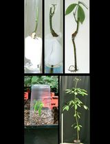
Figure 1. Required material and equipment. A. Five-day-old Arabidopsis seedlings. Scale bar: 1 cm. B. Materials needed to stain samples. C. Dissolving Fluorol Yellow 088 in lactic acid in a 50 ml Falcon tube attached to a vortex mixer with adhesive tape. D. Dissolved Fluorol Yellow (left) and aniline blue (right) solutions.
Equipment
Growth chamber to grow plant materia (Percival, CLF, Plant Climatics, AR75-L3 )
Vortex mixer (Instrumentfirman Labora, model: Vortex-Genie 2 )
Water bath (Fisher Scientific)
Soft stainless steel tweezers (3B DISSECTION SOFT TWEEZERS, LABWORLD)
Inverted confocal microscope (Zeiss, model: LSM880 )
Note: A fully motorized X, Y, Z scanning stage is required to perform tile scan image acquisition experiments.
Objectives: 10× or 20× (suitable for monitoring the whole specimen or root, e.g., Figure 3C). For higher resolution images, a higher magnification objective, such as 40× or 63×, is required (Figure 3D)
Fluorescence signal detection system for GFP and other fluorescent reporters (Shaner et al., 2007)
Software
Software operating the confocal microscope
ImageJ (Abràmoff et al., 2004)
Microsoft Excel
Prism (GraphPad)
Procedure
文章信息
版权信息
© 2021 The Authors; exclusive licensee Bio-protocol LLC.
如何引用
Marhavy, P. and Siddique, S. (2021). Histochemical Staining of Suberin in Plant Roots. Bio-protocol 11(3): e3904. DOI: 10.21769/BioProtoc.3904.
分类
植物科学 > 植物发育生物学 > 综合
分子生物学 > DNA > DNA 克隆
您对这篇实验方法有问题吗?
在此处发布您的问题,我们将邀请本文作者来回答。同时,我们会将您的问题发布到Bio-protocol Exchange,以便寻求社区成员的帮助。