

Induction of Epithelial-mesenchymal Transition in MDCK II Cells
诱导MDCK II细胞上皮-间充质转化

发布: 2021年02月05日第11卷第3期 DOI: 10.21769/BioProtoc.3903 浏览次数: 3897
评审: Savita NairIrit AdiniGiovanna Piovani
参见作者原研究论文
The authors used this protocol in:
Jun 2014


Abstract
Epithelial-mesenchymal transition (EMT) is a reversible process of epithelial cell transdifferentiation into a mesenchymal cell, that enables initiation of cell migration. EMT plays an important role in embryonic development, tissue repair and cancer metastasis. Better understanding of cellular and molecular events during EMT will not only provide novel insights on how mammalian organism develops and how epithelial tissues regenerate, but also can identify novel therapeutic targets for cancer therapy. Here we aim to provide a detailed protocol on how to induce EMT in Madin-Darby Canine Kidney (MDCK) II epithelial cell line and perform immunofluorescent staining on EMT-induced cells.
Keywords: Epithelial-mesenchymal transition (上皮细胞间质转型)Background
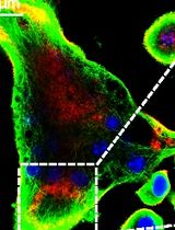
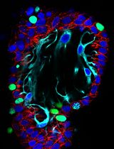
Epithelial cells are characterized by cell plasticity, that is the ability to adopt different cellular phenotypes (Carter et al., 2019; Yuan et al., 2019). Epithelial-mesenchymal transition (EMT) is a form of epithelial cell plasticity. During EMT epithelial cells disrupt cell-cell junctions, lose their polarity and change their shape from squamous, cuboidal, or columnar into spindle-like and become migratory, thus gaining the properties of mesenchymal cells (Kalluri and Weinberg, 2009). EMT can be evaluated by immunostainings and measurement of expression levels of markers such as E-cadherin, ZO-1, vimentin, fibronectin and N-cadherin (Kalluri and Weinberg, 2009). The studies on EMT during the last decade centered mostly on the role of transcription factors e.g., Snail1/2, ZEB1/2, Twist and microRNAs (Gonzalez and Medici, 2014), cytokines like tumor necrosis factor-alpha (TNF-α) and interleukin-6 (IL-6) (Yadav et al., 2011; Li et al., 2012), and signaling mechanisms such as transforming growth factor beta (TGF-β) signaling and NF-κB signaling (Gonzalez and Medici, 2014; Song et al., 2014; Dongre and Weinberg, 2019). However, regulatory mechanisms of early stages EMT and the precise sequence of molecular events during EMT remain largely unknown.
Madin-Darby Canine Kidney (MDCK) epithelial cell line is a cellular model to study epithelial cell polarity and junctions (Dukes et al., 2011; Yonemura, 2014; Vidal-Quadras et al., 2017). MDCK II cell line is a strain, that originates from higher passage of parental MDCK cells (Dukes et al., 2011). MDCK II cells differ from parental MDCK cells in that MDCK II cells have leaky cell junctions. Importantly, MDCK cells can be cultured in a standard 2D cell culture system, as well as in a more physiologically relevant 3D cell culture system (Yonemura, 2014; Vidal-Quadras et al., 2017).
EMT can be easily stimulated in 2D cell cultures of MDCK II cells by treatment with hepatocyte growth factor (HGF), that decreases cell roundness, upregulates the expression of vimentin, and increases the cell distance to the first and the second neighbor cell (Farrell et al., 2014). Here we provide a detailed description of the procedure on how to stimulate EMT in 2D cell cultures of MDCK II cells. Additionally, we describe the procedure for immunostaining of EMT-induced cells for ZO-1, a tight junction protein, and vimentin, the classical EMT marker.
Materials and Reagents
Sterile Cell Culture Dish, 100 × 20 mm (Sarstedt, catalog number: 83.3902 )
Serological pipettes: 5 ml (Sarstedt, catalog number: 86.1253.001 ), 10 ml (Sarstedt, catalog number: 86.1254.001 ), 50 ml (Sarstedt, catalog number: 86.1256.001 )
Tube 15 ml (Sarstedt, catalog number: 62.554.502 )
Tube 50 ml (Sarstedt, catalog number: 62.547.254 )
1.5 ml microtubes (Sarstedt, catalog number: 72.690.301 )
Culture chambers: Lumox 8-well specimen slide detachable (Sarstedt, catalog number: 94.6150.801)
Note: The procedure also works with FalconTM Chambered Cell Culture Slides, 8 wells, (Corning, catalog number: 354118).
Laboratory bottle, borosilicate 3.3 glass, 50 ml (VWR, catalog number: 215-3261 )
Laboratory bottle, borosilicate 3.3 glass, 1,000 ml (VWR, catalog number: 215-1595 )
Aluminium foil
ARTTM Barrier Specialty Pipette Tips: 10 µl (Thermo Scientific, catalog number: 2140 ), 200 µl (Thermo Scientific, catalog number: 2770 ), 1,000 µl (Thermo Scientific, catalog number: 2279 )
Microscope slide holder
Cover slips Menzel Glaser 24 × 60 mm (ThermoFisher Scientific, catalog number: E-4137 )
Parafilm
MDCK II cell line (Merck/Sigma-Aldrich, catalog number: ECACC 00062107 )
Eagle's Minimum Essential Medium (EMEM), 500 ml (ATCC, catalog number: 30-2003 )
Fetal bovine serum (FBS), 500 ml (Gibco, catalog number: 10270-106)
Note: FBS does not require heat inactivation.
Dulbecco's phosphate-buffered saline (DPBS) 1×, no calcium, no magnesium, 500 ml (Gibco, catalog number: 14190-094 )
Trypsin-EDTA (0.25%), phenol red, 100 ml (Gibco, catalog number: 25200056 )
Recombinant Human HGF (HEK293 derived), 25 μg (Peprotech, catalog number: 100-39H )
Goat normal serum, 10 ml (Agrisera, catalog number: AS10 1548 )
Saponin, 10 g (Sigma-Aldrich, catalog number: S4521 )
Paraformaldehyde (PFA), 1 kg (Sigma-Aldrich, catalog number: 16005 )
Sodium chloride (NaCl), 1 kg (VWR Chemicals, catalog number: 27810.295 )
Potassium chloride (KCl), reagent grade, Reag. Ph Eur, 1 kg (Scharlau, catalog number: PO02001000 )
di-Sodium hydrogen phosphate dihydrate (Na2HPO4·2H2O), 1 kg (Scharlau, catalog number: SO03391000 )
Potassium dihydrogen phosphate (KH2PO4), 1 kg (Merck, catalog number: 1048731000 )
VECTASHIELD® Antifade Mounting Medium with DAPI, 10 ml (Vector Laboratories, catalog number: H-1200 )
Nail polish
Sodium hydroxide (NaOH), 1 kg (Merck, catalog number: 1064691000 )
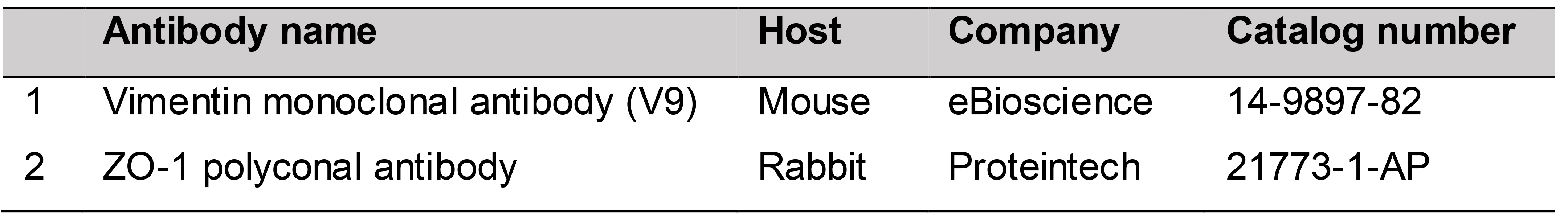
Primary antibodies (Table 1)
Table 1. Primary antibodies
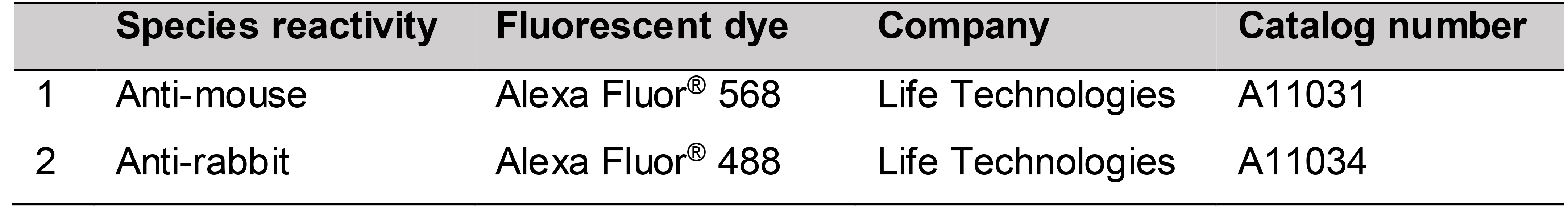
Secondary antibodies (Table 2)
Table 2. Secondary antibodies
Complete medium for MDCK II cells (see Recipes)
PBS 10× (see Recipes)
PBS 1× (see Recipes)
Blocking buffer (see Recipes)
Washing buffer (see Recipes)
3% PFA (see Recipes)
5% saponin (see Recipes)
Equipment
Counting chamber, type Burker (Hirschmann, catalog number: 8100201 )
Fume hood
Eppendorf® Research® plus pipette, 3-pack: 0.5-10 µl, 10-100 µl, 100-1,000 µl (Sigma-Aldrich, catalog number: Z683884 )
Integra Pipetboy 2 (VWR, catalog number: 612-0927 )
Heating and magnetic stirrer RH basic 2 (Dulova, catalog number: QSA337 )
Centrifuge VWR Compact Star CS4
37 °C, 5% CO2 cell culture incubator
Standard phase contrast microscope
Confocal microscope Zeiss 710
Software
Procedure
文章信息
版权信息
© 2021 The Authors; exclusive licensee Bio-protocol LLC.
如何引用
Pastuła, A. and Lundmark, R. (2021). Induction of Epithelial-mesenchymal Transition in MDCK II Cells. Bio-protocol 11(3): e3903. DOI: 10.21769/BioProtoc.3903.
分类
发育生物学 > 细胞生长和命运决定 > 分化
细胞生物学 > 基于细胞的分析方法
细胞生物学
您对这篇实验方法有问题吗?
在此处发布您的问题,我们将邀请本文作者来回答。同时,我们会将您的问题发布到Bio-protocol Exchange,以便寻求社区成员的帮助。