

Isolation of Extracellular Vesicles Derived from Mesenchymal Stromal Cells by Ultracentrifugation
超速离心法分离间充质基质细胞胞外囊泡


(*contributed equally to this work) 发布: 2020年12月20日第10卷第24期 DOI: 10.21769/BioProtoc.3860 浏览次数: 6503
评审: Xiaoyi ZhengMei Po ChanAlexandros C KokotosAnonymous reviewer(s)
参见作者原研究论文
The authors used this protocol in:

Jan 2020
Advertisement


Abstract
Extracellular vesicles (EVs) are a heterogeneous group of membranous vesicles that differ on their biogenesis and release pathways, such as exosomes, microvesicles and apoptotic bodies. They are involved in cell-to-cell communication delivering signal molecules (proteins, nucleic acids, lipids, etc.) that can regulate different physiological processes, as well as the development and progression of several diseases. There are different methods and commercial kits to isolate EVs and depending on the methodology one could obtain EVs with different degrees of efficiency, purity and it can be more or less time-consuming. Then, the choice has to be according to the different advantages and disadvantages, and their use for downstream applications. Here, we describe the EVs isolation method from mesenchymal stromal cells by ultracentrifugation. This EVs isolation can be performed using common media and buffers, and only with the requirement of an analytical ultracentrifuge. Moreover, this method can be used to obtain large quantity of EVs with a good reproducibility for developing in vitro and in vivo experiments and studying their biological actions.
Keywords: Mesenchymal stromal cells (间充质基质细胞)Background
Mesenchymal stem cells (MSCs) have a protective effect on the progression of different diseases contributing to the immunomodulation and the inflammatory state (Bartholomew et al., 2002; Togel et al., 2005; Azmi et al., 2013; Ebrahimi et al., 2013; Ben-Ami et al., 2014). Their protective action is not only due to their transdifferentiation, but also their paracrine mechanisms such as release of extracellular vesicles (EVs) showing an immunomodulatory, anti-inflammatory, anti-apoptotic and proangiogenic function (Bruno et al., 2009; Camussi et al., 2010a; Grange et al., 2020). Moreover, the application of EVs from MSCs in clinical practice has the advantage of being a safe therapy without the disadvantages related to other MSCs therapies, including the possibility to be rejected, gene stability, poor long-term differentiation and probability of viral transfer (Kunter et al., 2007; Wang et al., 2012).
In the last years, there is an increasing interest in the application of EVs in the transplant area, as an immunomodulatory and anti-inflammatory therapy on transplant recipients, and their addition on perfusion solution to pre-condition the graft before transplantation (Koch et al., 2015; Gregorini et al., 2017; Stone et al., 2017; Rigo et al., 2018; Ramírez-Bajo et al., 2020b). In particular, EVs therapy in the context of a hypothermic or normothermic perfusion machine could increase the long-term survival and increase the number of available organs. This improvement could be related to a decrease of inflammation, the preservation of energy metabolism and microvascular permeability avoiding the formation of edema. These properties show the great therapeutic potential of EVs in the transplantation process.
Cell-to-cell communication by EVs is a mechanism that has been preserved throughout the evolution in both prokaryotic and eukaryotic cells (Ratajczak et al., 2006). Since its discovery 30 years ago, it has been demonstrated that EVs are produced by an enormous variety of cell types such as blood cells, dendritic, endothelial and epithelial cells, nervous system cells, adult and embryonic stem cells and even tumor cells (Harding et al., 1984). EVs are a heterogeneous group of membranous vesicles that differ on their biogenesis and release pathways such as exosomes, microvesicles, microparticles, ectosomes, oncosomes and apoptotic bodies (Thery et al., 2002 and 2018). EVs act as paracrine/endocrine effectors that transport bioactive molecules among the cells (cytosolic proteins, lipids, mRNA, miRNA, DNA, mitochondrial DNA and genomic DNA) of the surrounding microenvironment, or being carried remotely in biological fluids (Fevrier and Raposo, 2004; Camussi et al., 2010b; Mittelbrunn et al., 2011). These regulatory biological signals can regulate various physiological processes, as well as the development and progression of disorders (Valadi et al., 2007; Guescini et al., 2010; Balaj et al., 2011).
A large number of studies are focused on EV’s field and their isolation from different biofluids, developing different methods of isolation such as ultracentrifugation, density gradient, size exclusion chromatography, filtration, microfluidics, precipitation kits and magnetic beads technologies. The choice of isolation methods should depend on the type of starting material, volume, available equipment, scientific inquiry and subsequent analysis or use (Momen-
Heravi et al., 2013). There are described different protocols associated with difference in purity and time of isolation. Moreover, EVs composition is dependent on the isolation protocol used, obtaining different subpopulations with different soluble proteins and nucleic acids. For these reasons, it is important to follow a series of criteria based on current best practice that represents the minimal experimental requirements for definition of EVs that allow the interpretation and their replication by other researchers (Lotvall et al., 2014).
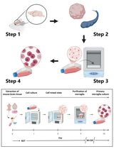
The most commonly used protocol for isolation is ultracentrifugation which has advantages such as the applicability for EVs isolation from large volumes as urine or cell culture conditioned media, the use of common media and buffers, easy to handle, and absence of impact on EVs except gravitational force (Konoshenko et al., 2018). This method can be used to obtain large quantity of EVs with a good reproducibility for developing studies about their biological actions. However, it needs expensive equipment and the isolated EVs could present co-isolating contaminants such as soluble proteins and nucleic acids. In our previous publications, we showed a comparison of the therapeutic effect of MSC and their EVs in an in vivo model of chronic kidney allograft rejection (Ramírez-Bajo et al., 2020a) and chronic cyclosporine nephrotoxicity (Ramírez-Bajo et al., 2020b). The ultracentrifugation protocol allows to produce a great quantity of EVs from high volumes of MSC-conditioned medium required for a periodic administration of therapies to different animal groups. We observed a good reproducibility between EVs batches by this methodology.
Materials and Reagents
22 G needle
15 ml conical tubes (Sarstedt, catalog number: 62.554.502 )
50 ml conical tubes (Sarstedt, catalog number: 62.548.004 )
Serological pipettes 10 ml (Sarstedt, catalog number: 86.1254.001 )
Serological pipettes 25 ml (Sarstedt, catalog number: 86.1685.001 )
Tissue Culture Flask, 150 cm2 filter (TPP, catalog number: 90151 )
Tissue Culture Flask, 75 cm2 filter (TPP, catalog number: 90076 )
0.22 μm sterile syringe filters (Sarstedt, catalog number: 831.826.001 )
0.1 μm sterile syringe filters (Pall Life Sciences, catalog number: 4668 )
20 ml Sterile syringes (BD Plastipak, catalog number: 10569215 )
Glass Pasteur pipettes (Deltalab, catalog number: 702 )
Countess cell counting chamber slides (Thermo Fisher Scientific, Invitrogen, catalog number: C10228 )
Open-Top Thinwall Ultra-Clear Tube, 38.5 ml capacity (Beckman Coulter, catalog number: 344058 )
Lewis and Fisher rats (200 g)
Trypan blue (Thermo Fisher Scientific, Invitrogen, catalog number: C10228 )
Alpha MEM (Lonza Bioscience, catalog number: BE02-002F )
RPMI 1640 Medium with L-Glutamine (Lonza Bioscience, catalog number: BE12- 702 F)
Fetal bovine serum (FBS) (EuroClone, catalog number: ECS0180L )
Penicillin-Streptomycin (10,000 U/ml) (Thermo Fisher Scientific, Invitrogen, catalog number: 15140122 )
Trypsin-EDTA (0.25%), phenol red (Thermo Fisher Scientific, Invitrogen, catalog number: 25200072 )
Dulbecco’s phosphate buffered saline (PBS) 10x, Without Ca++ and Mg++ (Cultek, catalog number: BE17-517Q )
Sterile water for irrigation, Serrasol solution (Serra Pamies, catalog number: 374561 )
Dimethyl sulfoxide (DMSO) (Sigma-Aldrich, catalog number: 276855 )
L-glutamine solution (Sigma-Aldrich, catalog number: G7513 )
Complete alpha-MEM medium (see Recipes)
Freezing medium (see Recipes)
Equipment
-80 °C freezer
Water bath set to 37 °C (Thermo Fisher Scientific, model: JP SelectaTM Precisterm 6000141 )
Refrigerated benchtop centrifuge (Thermo Fisher Scientific, model: HeraeusTM MultifugeTM X1)
Countess automated cell counter (Thermo Fisher Scientific, Invitrogen, catalog number: C10281 )
DHD Air-Jacketed Automatic CO2 Incubator (NuAire, catalog number: NU-5510E )
Inverted Modulation Contrast Microscope (Leica, model: DMIRB )
C2200 CBC Complete Balance (Cobos, 200-20018 )
Preparative ultracentrifuge (Beckman Coulter, model: Optima L100XP )
SW32 Ti Swinging-Bucket Rotor Package (Beckman Coulter, catalog number: 369694)
NanoDrop Microvolume Spectrophotometers (Thermo Fisher Scientific, model: ND1000)
NanoSight instrument equipped with a 638 nm laser and CCD camera (model F-033) (Malvern, model: LM10)
Cryo-electronmicroscope (Jeol, model: JEM 2011)
EMR Carbon support film on copper 400 square mesh (EMR, catalog number: 22-1MC040-100)
Flow cytometer (Becton Dickinson, Biosciences, model: FACS Canto II)
Software
FlowJo v10 software (BD Bioscience, https://www.flowjo.com/solutions/flowjo/downloads)
Nanosight NTA Software version 3.1 (build 3.1.46) (Malvern Panalytical, https://www.malvernpanalytical.com/en/learn/events-and-training/webinars/W150326NanoSightSoftwareRelease)
Procedure
文章信息
版权信息
© 2020 The Authors; exclusive licensee Bio-protocol LLC.
如何引用
Ramirez-Bajo, M. J., Banon-Maneus, E., Rovira, J., Campistol, J. M. and Diekmann, F. (2020). Isolation of Extracellular Vesicles Derived from Mesenchymal Stromal Cells by Ultracentrifugation. Bio-protocol 10(24): e3860. DOI: 10.21769/BioProtoc.3860.
分类
干细胞 > 成体干细胞 > 间充质干细胞
细胞生物学 > 细胞分离和培养 > 细胞分离
您对这篇实验方法有问题吗?
在此处发布您的问题,我们将邀请本文作者来回答。同时,我们会将您的问题发布到Bio-protocol Exchange,以便寻求社区成员的帮助。