- EN - English
- CN - 中文
Preparation of Bacterial Outer Membrane Vesicles for Characterisation of Periplasmic Proteins in Their Native Environment
细菌外膜囊泡的制备及其原生环境中周质蛋白的表征
发布: 2020年12月20日第10卷第24期 DOI: 10.21769/BioProtoc.3853 浏览次数: 4584
评审: David PaulElías Barquero-CalvoAnonymous reviewer(s)
Abstract
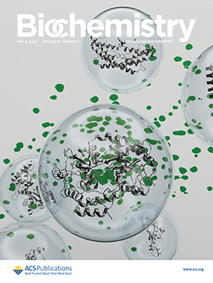
Bacterial outer membrane vesicles (OMVs) are naturally formed by budding from the outer membrane of Gram-negative bacteria. OMVs consist of a lipid bilayer identical in composition to the original outer membrane and contain periplasmic content within their lumen. Enriched with specific envelope proteins, OMVs make for an excellent native-like platform to study these proteins in-situ using biophysical methods. Here, we describe in detail the preparation of OMVs from Escherichia coli, which are luminally enriched with periplasmic proteins and uniformly labeled with stable isotopes (2H and 15N), suitable for the subsequent characterisation of proteins at atomic resolution in their native environment by solution-state NMR spectroscopy. The ability to perform structural studies of periplasmic components in-situ clears the way to reaching an in-depth understanding of the functional and mechanistic details of this unique cellular compartment.
Keywords: Outer membrane vesicles (外膜囊泡)Background
The periplasm of Gram-negative bacteria is a quite remarkable cellular compartment. This space, incarcerated between the inner and outer bacterial membrane, contains proteins at an extraordinarily high concentration exceeding 300 mg ml−1 (Oliver, 1996) and, in the absence of cellular sources such as ATP, functions almost energetically independent from its cytosolic counterpart. So far, structural knowledge about periplasmic proteins has been exclusively obtained using purified proteins isolated from their native environment. Thus, any structural and functional influence this special environment might impose on a protein are lost during purification. Efforts to study periplasmic proteins in-situ using biophysical methods such as in-cell NMR spectroscopy have been hampered by the low volume ratio of the periplasm, which contributes only 5-10% to the total bacterial volume (Brass et al., 1986).
We recently described a new method to characterize proteins within bacterial outer membrane vesicles (OMVs) using solution-state NMR spectroscopy (Thoma and Burmann, 2020). OMVs, which are naturally released by Gram-negative bacteria, contain periplasmic content in their lumen and can be non-destructively purified from bacterial growth cultures (Chutkan et al., 2013; Schwechheimer and Kuehn, 2015). Thus, OMVs represent an excellent platform to study periplasmic proteins, enriched in the luminal space, together with their natural environment. To be amendable for NMR studies, OMVs are uniformly labeled with stable isotopes (2H and 15N) and prepared in large quantities. Our approach proved particularly useful for the study of soluble periplasmic proteins within this environment, as the membrane components of OMVs remain largely invisible to solution-state NMR, due to slower molecular tumbling rates.
One key factor for obtaining large amounts of luminally enriched OMVs is the use of a suitable expression strain. Widely used expression hosts such as E. coli BL21(DE3) do not produce sufficient amounts of OMVs to be feasible for these types of experiments (Thoma et al., 2018). In contrast, strains lacking the major outer membrane protein OmpA have been shown to have a 26-fold increased OMV production (Schwechheimer et al., 2014). Moreover, deletion of OmpA omits possible background signals originating from its soluble peptidoglycan binding domain in NMR experiments (Thoma and Burmann, 2020). In our experimental set-up we relied on the strain BL21(DE3)ompA, which was kindly provided by Guido Grandi (Fantappiè et al., 2014) and which reproducibly yielded high amounts of luminally enriched OMVs. However, other alternatives such as BL21A are available (Meuskens et al., 2017) or could easily be tailor-made by deletion of the ompA gene in the expression strain of choice.
Another critical factor for obtaining large amounts of suitable OMVs is the time point, at which the bacteria are removed from the growth media. Bacterial growth should be maintained as long as possible, since the amount of OMVs present in the culture supernatant correlates directly with the cell density and consequentially with the length of the growth period. However, OMVs should be collected before bacterial growth ceases and cultures enter the stationary phase, because this will substantially increase contaminations with cytoplasmic proteins, inner membranes, and cell debris resulting from stress-induced explosive cell lysis (Turnbull et al., 2016; Thoma et al., 2018). Depending on whether cells are grown in H2O or D2O based media this stage is typically achieved after 3 to 5 h, respectively. Since bacterial growth is not only retarded by the use of D2O but also underlies various influences such as the strain and plasmid used for expression, it is advisable to determine the growth behavior of the bacterial cultures prior to OMV preparation. Whereas deuteration is not strictly necessary for small to medium sized proteins (< 25 kDa), luminally enriched OMVs prepared from cells grown in D2O based media typically yield NMR spectra of higher quality due to a decrease in background signals and a smaller NMR line-width (Thoma and Burmann, 2020).
Materials and Reagents
0.22 µm vacuum filtration unit (such as Steritop Quick Release, EMD Millipore, catalog number: S2GPT05RE )
0.45 µm vacuum filtration unit (such as Stericup-HV, EMD Millipore, catalog number: SCHVU11RE )
≥ 50 ml syringe (such as Fisherbrand Sterile Syringes, Thermo Fisher Scientific, catalog number: 15899152 )
0.45 µm syringe filter (such as Millex-HP, EMD Millipore, catalog number: SLHP033R )
Microscale NMR tubes (such as 5mm Shigemi Tube Set, Shigemi, catalog number: BMS-005B )
Tryptone (Sigma-Aldrich, catalog number: T9410 )
Yeast Extract (Sigma-Aldrich, catalog number: Y1625 )
NaCl (Sigma-Aldrich, catalog number: S7653 )
Na2HPO4 (Sigma-Aldrich, catalog number: S7907 )
KH2PO4 (Sigma-Aldrich, catalog number: P0662 )
KCl (Sigma-Aldrich, catalog number: P9333 )
CaCl2·2H2O (Sigma-Aldrich, catalog number: C5080 )
MgCl2·6H2O (Sigma-Aldrich, catalog number: M2670 )
15NH4Cl (Sigma-Aldrich, catalog number: 299251 )
D2O (Sigma-Aldrich, catalog number: 617385 )
D-Glucose (Sigma-Aldrich, catalog number: G7528 )
MgSO4 (Sigma-Aldrich, catalog number: M7506 )
Thiamine (Sigma-Aldrich, catalog number: T1270 )
Isopropyl-β-D-thiogalactoside (IPTG; Sigma-Aldrich, catalog number: I6758 )
LB-medium (Lennox) (see Recipes)
M9 medium (see Recipes)
Dulbeccos phosphate buffered saline with added salts (DPBSS)/NMR sample buffer (see Recipes)
Equipment
Shaking incubator (such as New Brunswick Innova® 42, Eppendorf AG, catalog number: M1335-0012 )
500 ml baffled culture flask (such as ROTILABO baffled flasks, Carl Roth, catalog number: LY96.1 )
5,000 ml baffled culture flask (such as ROTILABO baffled flasks, Carl Roth, catalog number: PK29.1 )
Cell density meter (such as GE Healthcare Ultrospec, Thermo Fisher Scientific, catalog number: 10704417 )
High speed centrifuge (such as Avanti J-series, Beckman Coulter, catalog number: B22989 )
High speed large-volume fixed angle rotor (such as J-LITE JLA-16.250, Beckman Coulter, catalog number: 363934) equipped with suitable centrifugation bottles (such as 250 ml Polycarbonate Bottle, Beckman Coulter, catalog number: 356013)
High speed small-volume fixed angle rotor (such as JA-25.50, Beckman Coulter, catalog number: 363055) equipped with suitable centrifugation tubes (such as 50 ml Polycarbonate Bottle, Beckman Coulter, catalog number: 357000)
Vacuum pump, sufficiently strong for use with vacuum filtration units (ultimate vacuum ~0.01 mPa)
Vortex mixer (such as Vortex-Genie 2, Scientific Industries, catalog number: SI-0246)
Microvolume spectrophotometer (such as Nanodrop One, Thermo Fisher Scientific, ND-ONE-W)
Gel electrophoresis system for SDS page (such as Mini-PROTEAN Tetra, Bio-Rad, catalog number: 1658004) including power supply, polyacrylamide gels, and necessary buffers and staining solutions (refer to manufacturers specifications for details)
Software
Any spreadsheed or scientific graphing software allowing basic calculations (Excel, Numbers, LabPlot, Origin, etc.)
Procedure
文章信息
版权信息
© 2020 The Authors; exclusive licensee Bio-protocol LLC.
如何引用
Thoma, J. and Burmann, B. M. (2020). Preparation of Bacterial Outer Membrane Vesicles for Characterisation of Periplasmic Proteins in Their Native Environment. Bio-protocol 10(24): e3853. DOI: 10.21769/BioProtoc.3853.
分类
生物化学 > 蛋白质 > 分离和纯化
微生物学 > 微生物生物化学 > 蛋白质
您对这篇实验方法有问题吗?
在此处发布您的问题,我们将邀请本文作者来回答。同时,我们会将您的问题发布到Bio-protocol Exchange,以便寻求社区成员的帮助。
Share
Bluesky
X
Copy link


