

A Quantitative Assay to Measure Stress Granule Association of Proteins and Peptides in Semi-permeabilized Human Cells
半透性人细胞中蛋白质和肽应激颗粒结合的定量测定
发布: 2020年12月20日第10卷第24期 DOI: 10.21769/BioProtoc.3846 浏览次数: 5695
评审: Steven BoeynaemsIndranil MalikAnonymous reviewer(s)
参见作者原研究论文
The authors used this protocol in:

Apr 2018


Abstract
Stress granules (SGs) are membrane-less organelles that form in the cytoplasm through phase separation, in response to diverse stressors. SGs contain translationally stalled mRNAs, proteins involved in translation, and various RNA-binding proteins (RBPs). Due to the high local concentration of aggregation-prone RBPs, SGs might act as condensation sites for aberrant phase transitions of RBPs and could favor formation of solid protein aggregates underlying the pathological cytoplasmic inclusions found in numerous neurodegenerative diseases. Most assays aiming at studying the recruitment of RBPs into SGs are based on overexpression and SG recruitment of RBPs in intact cells. These approaches are, however, often limited by the predominantly nuclear localization of many RBPs, which precludes cytoplasmic RBP concentrations sufficient for SG localization, and does not address RBP recruitment independent of SG formation. Here, we present a quantitative method to assess recruitment of recombinant RBPs into pre-formed SGs, independent of the RBP’s nuclear localization, using semi-permeabilized cells and fluorescence microscopy. In this assay, SGs are firstly induced by a stressor, and then the plasma membrane of the stressed cells is subsequently selectively permeabilized to provide access of the recombinant protein to SGs. Nuclear import of the protein-of-interest is prevented by blocking nuclear pores with wheat germ agglutinin. This assay allows one to study the molecular mechanisms underlying recruitment of RBPs into SGs quantitatively, in absence of their nuclear import and under controlled conditions. The method allows for a direct comparison of wildtype, mutant or posttranslationally modified RBPs, for addressing the influence of other proteins’ preventing or promoting SG association of RBPs, and is also applicable to synthetic peptides.
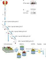
Graphic abstract
Workflow overview for analysis of SG recruitment of recombinant proteins or peptides in semi-permeabilized cells
Background
Stress granules (SGs), membrane-less organelles that contain translationally stalled mRNAs and several RNA-binding proteins (RBPs), form by phase separation as part of a cytoprotective mechanism in response to cellular stress (Protter and Parker, 2016; Gomes and Shorter, 2019). However, SGs have also been suggested to act as precursors for pathological protein aggregates in neurodegenerative diseases, in particular amyotrophic lateral sclerosis (ALS) and frontotemporal dementia (FTD) (Bentmann et al., 2013; Alberti and Dormann, 2019; Wolozin and Ivanov, 2019; Zhang et al., 2019). First, pathological inclusions containing the RBPs Fused in Sarcoma (FUS) or TAR DNA-binding protein 43 (TDP-43) in ALS/FTD post mortem tissue have been shown to harbor proteins commonly found in SGs, such as PABPC1 or TIA-1 (Dormann et al., 2010; Liu-Yesucevitz et al., 2010; Bentmann et al., 2012; Mackenzie et al., 2017). Second, ALS-associated mutations in RBPs, such as FUS, TDP-43, several hnRNPs and TIA-1, were demonstrated to promote SG localization of these proteins and/or to reduce SG dynamics (Bosco et al., 2010; Dewey et al., 2011; Dormann et al., 2010; Kim et al., 2013; Mackenzie et al., 2017). Third, optogenetic approaches have elegantly demonstrated that persistent or repetitive formation of SGs can lead to cytoplasmic assemblies resembling pathological inclusions (Zhang et al., 2019). Disease-linked RBPs have the tendency to phase separate at high concentration in vitro into liquid-like condensates that can solidify over time, a process promoted by distinct ALS-associated mutations (Murakami et al., 2012; Molliex et al., 2015; Patel et al., 2015; Mackenzie et al., 2017). As SGs contain a high local protein concentration, they could act as condensation sites for disease-linked RBPs and thus promote their pathological aggregation.
What are the molecular mechanisms that determine SG association of RBPs and other proteins? In the past years, the role of individual domains or mutations of RBPs for SG localization was mainly studied by cellular (over) expression of RBPs. However, this approach does not distinguish (i) whether the ectopically expressed protein might promote or suppress the process of SG formation per se, (ii) whether reduced nuclear import of the protein-of-interest might increase its cytoplasmic concentration, and thereby its partitioning into SGs, or (iii) whether a specific mutation directly promotes or suppresses SG association of the protein-of-interest.
Here, we report an assay to study association of recombinant proteins or peptides with pre-formed SGs in semi-permeabilized cells by quantitative fluorescence microscopy. It has been adapted from an original protocol by Adam et al. (1992) who first established selective permeabilization of the plasma membrane by the detergent digitonin to study mechanisms of nuclear import of proteins. Our assay can determine SG association of recombinant proteins or peptides irrespective of their nuclear import, as nuclear pore complexes are selectively blocked by wheat germ agglutinin (WGA), known to bind to nucleoporins (Finlay et al., 1987). By the use of recombinant proteins or peptides, either as fusion protein or untagged, SG association in dependence of the protein sequence or post-translational modifications can directly be correlated with further biochemical characterization in vitro (Hofweber and Dormann, 2018; Ukmar-Godec et al., 2019; Bourgeois et al., 2020). Furthermore, the influence of other factors, such as molecular chaperones or interacting proteins, can be studied under controlled conditions in a quantitative manner (Hofweber and Dormann, 2018; Bourgeois et al., 2020). This protocol was first published by Hofweber and Dormann (2018) to study how SG association of FUS is altered by its nuclear import receptor and chaperone Transportin (TNPO1) as well as arginine methylation.
Materials and Reagents
High precision coverslips, 12 mm No 1.5 (Marienfeld, catalog number: 0 117520 )
Microscopy slides, 76 x 26 mm (any commercial available, e.g., Marienfeld, catalog number: 1000200 )
6- or 10-cm cell culture dishes (any commercially available, e.g., Thermo Fisher Scientific, catalog number: 150288 or Greiner Bio-One, catalog number: 664160 , respectively)
Humid-chamber (e.g., wet Whatman paper covered by parafilm in a 10- or 15 cm cell culture dish) (sterile)
Custom made recombinant protein or peptide (either fluorescently labelled or tagged suitably for immunostaining)
Milli-Q water
HEPES (any commercially available, e.g., Sigma, catalog number: H3375 )
Potassium acetate (any commercially available, e.g., Roth, catalog number: T874.2 )
Magnesium acetate (any commercially available, e.g., MERCK, catalog number: 7463348 )
EGTA (any commercially available, e.g., GERBU, catalog number: 1310 )
Potassium phosphate monobasic (KH2PO4) (any commercially available, e.g., Roth, catalog number: 3904.1 )
Potassium phosphate dibasic (K2HPO4·3H2O) (any commercially available, e.g., Roth, catalog number: 6878.1 )
Magnesium chloride (MgCl2) (any commercially available, e.g., Sigma, catalog number: M2670 )
Sodium chloride (any commercially available, e.g., Roth, catalog number: 0 601.2 )
Potassium chloride (KCl) (any commercially available, e.g., Roth, catalog number: 6781.1 )
Disodiumhydrogen phosphate (Na2HPO4) (any commercially available, e.g., Roth, catalog number: P030.2 )
DMSO (Sigma, catalog number: D2438 )
MG-132 (10 mM in DMSO) (Sigma, catalog number: C2211 )
DMEM high glucose (Thermo Fisher Scientific, Invitogen, catalog number: 61995059 )
10x Trypsin-EDTA (Sigma, catalog number: 59418C )
Sterile PBS solution (see solutions)
Fetal bovine serum (FBS) (Thermo Fisher Scientific, Invitogen, catalog number: 10270-106 )
Gentamycin (e.g., Thermo Fisher Scientific, Invitogen, catalog number: 15710049 )
Digitonin, high purity (Milipore, CAS: 11024-24-1 )
Trypan Blue solution (Thermo Fisher Scientific, Invitogen, catalog number: 152 500 61 )
Wheat germ agglutinin (WGA) (lectin from Triticum vulgaris) solution (Sigma, catalog number: A2408 )
37% Formaldehyde (AppliChem, catalog number: 131328.1211 )
TX-100 (any commercially available, e.g., AppliChem, catalog number: 9002-93-1 )
Serum matching the species that the secondary antibodies was raised in, e.g., donkey serum (Milipore, catalog number: S30 )
Tween-20 (Roth, catalog number: 9127.2 )
DAPI (Sigma, catalog number: D9542 )
ProLong Diamond Antifade mountant (Thermo Fisher Scientific, Invitogen, catalog number: P36961 )
DL-Dithiothreitol (DTT, Applichem, catalog number: A1101.0025 )
Aprotinin (Roth, catalog number: A162.2 )
Leupepstin hemisulfate (Roth, catalog number: CN33.2 )
Pepstatin A (Roth, catalog number: 3083.2 )
0.1% (w/v) poly-L-lysine solution (Sigma, catalog number: P8920 )
Antibody to detect SG core components, e.g., Rabbit anti-G3BP1 antibody (ProteinTech, catalog number: 13057-2-AP )
Optional: Antibody to detect tag of recombinant protein, e.g., Mouse anti-MBP antibody (ProteinTech, catalog number: 66003-1-1g )
Optional: Protein labeling kit (any commercially available, e.g., Thermo Fisher Scientific, Invitogen, catalog number: 46403 )
Fluorescently labelled secondary antibodies (e.g., donkey anti-rabbit Alexa 555 or Alexa 647; Thermo, catalog number: A-31572 or A-31573 ; donkey anti-mouse A-21202)
DMEM/10% FBS/10 µg/ml gentamycin (see Recipes)
1x Trypsin/EDTA (see Recipes)
10 mM MG-132 in DMSO (see Recipes)
Reaction buffer, either transport buffer (TPB) or potassium-phosphate buffer (KPB) (see Recipes)
10% digitonin/reaction buffer (see Recipes)
2 mg/ml WGA in reaction buffer (see Recipes)
3.7% formaldehyde in PBS (see Recipes)
10% and 0.5% TX-100/PBS solution (see Recipes)
PBS/0.1% Tween-20 (see Recipes)
1% donkey serum in PBS/0-1% Tween or alternative blocking solution for immunostaining (see Recipes)
DAPI solution (see Recipes)
Equipment
P1000, P200, P20, P10 pipettes
Fine tip curved tweezers
Cell culture hood
CO2 incubator
pH meter
Vacuum pump
Optional: upright microscope for brightfield microscopy
Confocal scanning microscope (e.g., Leica SP8 with 63x/1.4 oil objective or equivalent) equipped with laser lines matching chosen secondary antibodies and protein/peptide labels. Detection by standard photomultiplier tube (PMT) has been sufficient for visualization and quantification of protein localization to SGs in our hands.
Software
LASX software (Leica)
Fiji/ImageJ (NIH, https://imagej.net/Fiji)
Excel (Microsoft)
GraphPad Prism (GrapPad)
Procedure
文章信息
版权信息
© 2020 The Authors; exclusive licensee Bio-protocol LLC.
如何引用
Hutten, S. and Dormann, D. (2020). A Quantitative Assay to Measure Stress Granule Association of Proteins and Peptides in Semi-permeabilized Human Cells. Bio-protocol 10(24): e3846. DOI: 10.21769/BioProtoc.3846.
分类
神经科学 > 细胞机理
细胞生物学 > 基于细胞的分析方法 > 蛋白互作
生物化学 > 蛋白质 > 相互作用 > 蛋白质-配体相互作用
您对这篇实验方法有问题吗?
在此处发布您的问题,我们将邀请本文作者来回答。同时,我们会将您的问题发布到Bio-protocol Exchange,以便寻求社区成员的帮助。