

Acidified Blue Ink-staining Procedure for the Observation of Fungal Structures Inside Roots of Two Disparate Plant Lineages
两个不同植物谱系根内真菌结构的酸化蓝墨水染色法观察流程


.JPEG)
(*contributed equally to this work) 发布: 2020年10月20日第10卷第20期 DOI: 10.21769/BioProtoc.3786 浏览次数: 5115
评审: Yueqiang LengAnonymous reviewer(s)
参见作者原研究论文
The authors used this protocol in:
Jul 2020
Advertisement


Abstract
Identifying microscopic mycorrhizal fungal structures in roots, i.e., hyphae, vesicles and arbuscules, requires root staining procedures that are often time consuming and involves chemicals known to present health risks from exposure. By modifying established protocols, our root staining method stains roots using a safe ink- and vinegar-based staining solution, followed by a 2-16 h-long de-staining period. The entire procedure can be completed in less than 6 h (plus up to 16 h de-staining overnight) and roots are suitable for semi-permanent and permanent slide mounting for light microscopy. We tested our method on hundreds of wild-sourced roots from two different plant species: Lycopodiella inundata, a herbaceous clubmoss with tough water-resistant roots, and Sambucus nigra, a temperate woody shrub. Both plants associate with endomycorrhizae, L. inundata predominantly with Mucoromycotina fine root endophytes (MucFRE) and S. nigra with Glomeromycota arbuscular mycorrhizal fungi (AMF). Here we describe a simple, efficient, repeatable and safe method to detect the presence of fungal structures using light microscopy.
Keywords: Fine root endophytes (细根内生菌)Background
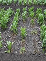
Here we detail an efficient and safe staining method used to analyse 1,305 Lycopodiella inundata (Figure 1A) roots for fine root endophytes (FRE) (Kowal et al., 2020) and 144 Sambucus nigra roots for coarse or AMF. We also describe how to clean roots for staining and prepare semi-permanent slides to analyse the presence/absence of these two widespread mycorrhizas present in host plant roots across the land plant phylogeny.
Established staining methods for identifying mycorrhizal fungal structures typically used trypan blue or chlorazol black E dyes (Phillips and Hayman, 1970; Agerer, 1991; Brundrett et al., 1994), understood to be carcinogenic. Other researchers described a safer alternative using an ink-vinegar staining solution and clearing with KOH (Vierheilig et al., 1998 and 2005; Wilkes et al., 2019) and compared techniques (Vierheilig et al., 2005), but none tested these techniques with significantly finer FRE (hyphae < 2 μm wide) as observed in Lycopodiella inundata, a lycopod. We demonstrate this same method is reliable and repeatable in host plant from a later evolved angiosperm lineage, Sambucus nigra, which is colonised by wider coarse hyphae and AMF.
Materials and Reagents
- Root cleaning
- Root material/plant
- Ethanol (EtOH) (Fisher Scientific)
- Distilled water (dH2O)
- Root clearing and staining
- Non-filter pipette tips (SARSTEDT)
- 2 ml microcentrifuge tubes (Eppendorf)
- Distilled water (dH2O)
- Blue ink (Sheaffer, catalog number: 94221 )
- Glycerol (Sigma-Aldrich)
- Glacial acetic acid (Sigma-Aldrich)
- Acetic acid (C2H4O2) (Sigma-Aldrich)
- Lactic acid (Sigma-Aldrich)
- Potassium hydroxide (KOH) (Fisher Scientific)
- Staining solution–10% Sheaffer blue ink in 25% glacial acetic acid (see Recipes)
- De-staining solution 1–1% acetic acid (see Recipes)
- De-staining solution 2–50% glycerol (see Recipes)
- Microscopy
- Glass slides 76 x 26 mm/0.8-1.0 mm (Thermo Fisher Scientific) and cover slips 18 x 18 mm/0.16-0.19 mm (VWR)
- Clear nail polish
- Mounting solution–50% glycerol for semi-permanent slides, or PVLG (Polyvinyl-Lacto-Glycerol) for permanent slides (see Recipes for both)
Equipment
- Root cleaning
- Fridge or cool box
- Wash bottle
- Paint brush
- Small plastic dish with shallow sides
- Airtight vessels for storing cleaned roots (Falcon tubes or glass jars are also suitable)
- Blunt forceps
- Root clearing and staining
- Scalpel blade
- Blunt forceps
- Wash bottle
- 2 microcentrifuge tube racks (1 of which should be heat proof)
- Water bath (we used Nickel-Electro Ltd–Clifton)
- Washing tray containing individual shallow wells (can be glass, plastic, metal or ceramic)
- Plastic tray
- Pipettes
- Microscopy
- Stereomicroscope (Leica, model: MZ6 ) with halogen cold light source (Volpi, model: Intralux-4001 ). Objectives required: 10x, 20x, 40x, 63x (MucFRE only)
- Light microscope (Leica, model: DMLB ) coupled to a camera (Leica, model: DMC5400 )
- Sewing needle or pin
- Warming oven (only if PVLG mounting solution used for permanent slides)
Software
- Leica Application Suite (LAS) imaging software version 4.13 (Leica)
Procedure
文章信息
版权信息
© 2020 The Authors; exclusive licensee Bio-protocol LLC.
如何引用
kowal, J., Arrigoni, E. and Lane, S. (2020). Acidified Blue Ink-staining Procedure for the Observation of Fungal Structures Inside Roots of Two Disparate Plant Lineages. Bio-protocol 10(20): e3786. DOI: 10.21769/BioProtoc.3786.
分类
植物科学 > 植物细胞生物学 > 细胞染色
微生物学 > 微生物-宿主相互作用 > 真菌
细胞生物学 > 细胞染色 > 全细胞
您对这篇实验方法有问题吗?
在此处发布您的问题,我们将邀请本文作者来回答。同时,我们会将您的问题发布到Bio-protocol Exchange,以便寻求社区成员的帮助。