

Evaluation of Viable Cells in Pseudomonas aeruginosa Biofilms by Colony Count and Live/Dead Staining
菌落计数和活/死染色法评价铜绿假单胞菌生物膜中的活细胞

发布: 2020年09月20日第10卷第18期 DOI: 10.21769/BioProtoc.3762 浏览次数: 6873
评审: Juan Facundo Rodriguez AyalaAlba BlesaChao Jiang
参见作者原研究论文
The authors used this protocol in:
Jan 2020
Advertisement


Abstract
Pseudomonas aeruginosa is a human pathogen capable to form robust biofilms. P. aeruginosa biofilms represent a serious problem because of the adverse effects on human health and industry, from sanitary and economic points of view. Typical strategies to break down biofilms have been long used, such as the use of disinfectants or antibiotics, but also, according to their high resistance to standard antimicrobial approaches, alternative strategies employing photocatalysis or control of biofilm formation by modifying surfaces, have been proposed. Colony forming units (cfu) counting and live/dead staining, two classic techniques used for biofilm quantification, are detailed in this work. Both methods assess cell viability, a key factor to analyze the microbial susceptibility to given treatment, then, they represent a good approach for evaluation of an antibiofilm strategy.
Keywords: Pseudomonas aeruginosa (铜绿假单胞菌)Background
Bacterial biofilms, complex structures attached to surfaces, are matrices composed by proteins, DNA, polysaccharides and water networks, in which cells are embedded (Costerton et al., 1995). Pseudomonas aeruginosa is a versatile bacterium that can be found in terrestrial and aquatic environments, or as human pathogen, either as free cells or as cells in robust biofilms. P. aeruginosa biofilms represent a serious problem because of the adverse effects on human health and industry (Nickel et al., 1985; Gibson et al., 1999; Willcox et al., 2001; Ramsey and Wozniak, 2005; Rajasekar et al., 2010; Mulcahy et al., 2014) and their high resistance to antibacterial agents (Mah and O’Toole, 2001; Mah et al., 2003). Because of their resistance and robustness, P. aeruginosa biofilms represent a model for biofilm studies (Ciofu and Tolker-Nielsen, 2019). The development of P. aeruginosa biofilms is regulated by a complex genetic program; in addition, biofilm formation is modified by environmental factors (O’Toole et al., 2000; Di Bonaventura et al., 2007; Ben Said et al., 2011; Gambino and Cappitelli, 2016; Pezzoni et al., 2018).
Factors related to the high resistance of biofilms include impaired diffusion of antibacterial compounds, reduced sensitivity due to slow growth rate of cells in biofilms, emergence of resistant bacterial phenotypes, presence of antioxidant products in the biofilm matrix, among others (Pezzoni et al., 2014; Hall and Mah, 2017). Strategies employed to combat biofilms include chemical and physical treatments such as application of disinfectants, antibiotics and ultrasound (Bridier et al., 2011; Wu et al., 2014; Gnanadhas et al., 2015). The high resistance of biofilm cells to commonly used disinfectants and the risk to human health and the environment by the use of increasing bactericide doses prompted to redirect research to safer strategies, such as the use of photocatalytic techniques, proposed as inexpensive, safe and effective (Dalrymple et al., 2010; Gamage McEvoy and Zhang, 2010; Pezzoni et al., 2020). On the other hand, since adhesion of microorganisms to surfaces depends on the surface topography and roughness, preventing biofilm formation by tuning surfaces at nanoscale level is a major opportunity in the development of antibiofilm strategies (Katsikogianni and Missirlis, 2004; Pezzoni et al., 2017).
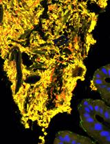
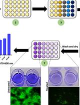
In order to deepen in the understanding of biofilm properties and/or evaluate the effectiveness of antibacterial treatments, several techniques have been employed to evaluate the amount of live bacteria in biofilms (Welch et al., 2012). Among them, two known methods are the counting of the number of colony forming units (cfu) and the live/dead staining (Merritt et al., 2005; Smith and Hunter, 2008; Tsvetanova, 2020). Both methods provide valuable but different information. In the cfu method, a viable cell is one capable to form a colony. On the other hand, the live/dead staining evaluates membrane integrity. The live/dead staining method employs the fluorescent stains SYTO 9 and Propidium iodide (PI), and viability is evaluated by fluorescence microscopy. PI is a red dye that only enters cells with permeabilized cytoplasmic membrane, while SYTO 9 is a green dye that stains all types of cells. According to this criterion, green cells (intact membrane) are live and red cells (disrupted membrane) are dead. While the colony count method requires biofilm disruption and a subsequent step of cell culture to visualize individual colonies, the live/dead method can be applied on entire biofilms and allows us to evaluate the morphological aspects of the biofilm. Since the two methods are based on different criteria, the data obtained from them will not be identical but they will follow the same tendency upon a given antibacterial treatment (Berney et al., 2006; Bosshard et al., 2010; Pezzoni et al., 2014 and 2020). In this work, we detailed how to apply both methods to P. aeruginosa biofilms.
Materials and Reagents
- 14 ml sterile Borosilicatetubes (Pyrex®, catalog number:SLW1622/09M))
- 1.5 ml sterile Eppendorf centrifuge tubes (Eppendorf, catalog number: 022364111)
- Sterile pipette tips 1-200, 100-1,000 (Corning®, catalog numbers: S4860, S9032)
- Sterile Petri dish glass plates (40 mm and 80 mm diameter) (Brand®, catalog numbers: BR455701, BR455732)
- Sterile cotton plugs (Nunn Finer, catalog number: 562)
- Glass coupons (approximately 15 mm x 15mm x 1mm)
Obtained from glass slides cut with an emery stone. To prepare the coupons, the slides are sliced gently with successive passes of the emery stone. You can wet the area to facilitate the cut. Finally, the glass slide is wrapped with a towel and the final cut is made by pressing hard on the marked area. - Glass slides (YEGREN®, catalog number: 7101)
- Emery stone (Value-Tec, catalog number 52-003094)
- Tupper container (20 cm x 30 cm x 6 cm)
- Rake shaped glass spreaders (Chemglass Life Science, catalog number: CLS-1350-01)
- Aluminum foil (Reynolds Wrap Heavy Duty Aluminum Foil, catalog number: 625)
- Paper towel (WypAll* X 60 Jumbo Roll, KCWW, Kimberly-Clark, catalog number: 30218593)
- Inoculating loop (DeconTM, catalog number: MP 19025)
- P. aeruginosa strain PAO1 (B.H. Holloway)
Pseudomonas aeruginosa PAO1 is maintained in glycerol stocks in liquid nitrogen. Stocks are prepared by mixing by inversion 130 µl of sterile glycerol and 770 µl of an overnight bacterial culture grown in LB medium. Bacteria are streaked with a sterile inoculating loop from glycerol stocks onto solid LB plates and grow overnight at 37 °C in an incubation stove. Single colonies are used as inoculums. - Glycerol (Sigma-Aldrich, catalog number: G5516-500ML)
- Distilled water (G-BIOSCIENCES, catalog number: 786-1713)
- Etanol 96% (EMSURE® Reag, catalog number: 159010)
- Inmersion oil (Leica, standar and type “F”)
- Tryptone (OXOID, catalog number: LP0042)
- Yeast extract (Merck, catalog number: 103753)
- Granulated agar (Difco, catalog number: 214530)
- Sodium Chloride (NaCl) (Biopack, catalog number: 1646.08)
- SYTOTM 9 Green Fluorescent Nucleic Acid Stain (Invitrogen, catalog number: S34854)
- Propidium Iodide (Invitrogen, catalog number: P1304MP)
- LB medium (see Recipes)
- LB agar solid medium (see Recipes)
- 4 M NaCl solution(see Recipes)
- Saline solution (see Recipes)
- 3.5 μM SYTO 9 and 20 μM PI solution (see Recipes)
Equipment
- 125 ml sterile Erlenmeyers flasks (Duran®, catalog number: 2121628)
- 2-20 µl, 20-100 µl,100-1,000 µl Kartell pluripet micropipettes (Kartell LABWARE, catalog numbers: 13000, 13210, 13220) and 1-10 ml Acura® manual micropipette (Socorex Swiss, catalog number: 825/835)
- Sterile 50, 100 and 1,000 ml borosilicate measuring cylinders (VILABO, catalog numbers: 3501114, 3501115, 3501118)
- Sterile 100 ml glass beakers (Brand®, catalog number: BR91224)
- Spatula (Arnaldochapini®, catalog number: 1929)
- Stainless steel dissecting tweezer (Fisherbrand, catalog number: 12-000-132)
- Conventional incubator shaker (New Brunswick Scientific Co., INC, model: G25)
- UV-Vis Spectrophotometer (Biotraza, model: 752)
- Autoclave (Hirayama HICLAVETM, model: HVE-50)
- Hot air oven sterilizer (DalvoIntrumentos, model: OHR/T)
- Vortex (Velp, model: ZV3, 201251076)
- Epifluorescence microscope (Olympus, model: BX51)
- Incubation stove (Precision, Scientific Group, model 4, catalog number: 31483)
- Bunsen burner (EiscoTM, catalog number: CH0091B)
Software
- ImageJ software (Rasband, 1997)
Procedure
文章信息
版权信息
© 2020 The Authors; exclusive licensee Bio-protocol LLC.
如何引用
Pezzoni, M., Pizarro, R. A. and Costa, C. S. (2020). Evaluation of Viable Cells in Pseudomonas aeruginosa Biofilms by Colony Count and Live/Dead Staining. Bio-protocol 10(18): e3762. DOI: 10.21769/BioProtoc.3762.
分类
微生物学 > 微生物生物膜 > 生物膜培养
细胞生物学 > 细胞活力 > 细胞增殖
您对这篇实验方法有问题吗?
在此处发布您的问题,我们将邀请本文作者来回答。同时,我们会将您的问题发布到Bio-protocol Exchange,以便寻求社区成员的帮助。