

Candida albicans Agar Invasion Assays
白念珠菌琼脂侵入实验

发布: 2020年08月20日第10卷第16期 DOI: 10.21769/BioProtoc.3730 浏览次数: 5555
评审: Kristin L. ShinglerChao JiangAnonymous reviewer(s)
参见作者原研究论文
The authors used this protocol in:
Nov 2019
Advertisement


Abstract
The ability of the human fungal pathogen Candida albicans to disseminate into tissues is promoted by a switch from budding to invasive hyphal growth. This morphological transition is stimulated by multiple environmental factors that can vary at different sites of infection. To identify genes that promote invasive growth, C. albicans mutants can be screened for defects in growing invasively into solid agar medium as a substitute for studying tissue invasion. This in vitro approach has advantages in that it permits the media conditions to be varied to mimic different host environments. In addition, the concentration of agar can be varied to determine the effects of altering the rigidity of the matrix into which the cells invade, as this provides a better indicator of invasive growth than the ability to form hyphae in a liquid culture. Testing under multiple conditions can be used to identify mutant cells with the strongest defects. Therefore, protocols and media for analyzing invasive growth of C. albicans under different conditions will be described that are appropriate for testing a single strain or high-throughput analysis of a collection of mutant C. albicans strains.
Keywords: Hyphae (菌丝)Background
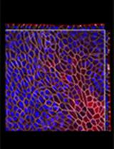
Candida albicans is a multimorphic fungal pathogen that can grow by forming buds (small spherical cells), pseudohyphal cells (chains of elongated cells), or hyphal cells (long filamentous chains of cells with parallel walls) (Figure 1) (Noble et al., 2017). A wide range of environmental stimuli encountered in the host have been shown to promote the switch to invasive growth, including human body temperature (37 °C), alkaline pH, CO2, factors in serum, and the sugar GlcNAc (N-acetyl glucosamine) (Kornitzer, 2019). Budding cells are thought to facilitate bloodstream dissemination and colonization of the gastrointestinal tract (Pierce and Kumamoto, 2012; Witchley et al., 2019). In contrast, the filamentous pseudohyphal and hyphal forms of C. albicans are better able to promote invasive growth into tissues (Noble et al., 2017). For example, our recent studies have shown a key role for invasive growth in establishing oral C. albicans infections (Naseem et al., 2019).
Genetic screens have identified a large number of mutants that regulate the ability of C. albicans to switch from budding to hyphal growth (Noble et al., 2010; O'Meara et al., 2015; Glazier et al., 2017; Kornitzer, 2019). However, a weak hyphal stimulus was often used in these studies as a way to sensitively detect a partial defect. Many of the previously reported mutants can still be induced by stronger stimuli, such as those encountered in the host (Riggle et al., 1999; Noble et al., 2010; Naseem et al., 2011; Douglas et al., 2013). Other mutants are only defective in forming hyphae in liquid medium and can still invade into a solid substrate. This phenotype is likely caused by contact with a solid substrate, which triggers an independent hyphal pathway (Kumamoto, 2005 and 2008; Douglas et al., 2013). Therefore, to identify genes that are broadly important for invasive hyphal growth, we screened for C. albicans mutants that were defective in invading into solid agar media under a diverse set of strongly inducing conditions (Naseem et al., 2019). Analysis of the strongest mutants in a mouse model of oral candidiasis revealed an important role for the RVS161 and RVS167 genes, which are known to promote the scission phase of endocytosis (Naseem et al., 2019). The RVS genes are also important for systemic infections caused by C. albicans, further implicating endocytosis in virulence (Douglas et al., 2009).
Blocking endocytosis may have therapeutic value, as small molecules inhibitors of endocytosis and hyphal growth showed efficacy against oral candidiasis (Pierce et al., 2015; Bar-Yosef et al., 2017; Romo et al., 2017; Vila et al., 2017). Furthermore, the rvs and other C. albicans endocytosis mutants were much more susceptible to killing by copper (Douglas and Konopka, 2019), suggesting copper could enhance the therapeutic value of endocytosis inhibitors. Thus, C. albicans agar invasion assays can be useful for screening mutant strains and novel inhibitors for efficacy in blocking the invasive growth of C. albicans.

Figure 1. Morphologies of C. albicans cells. Scale bar, 5 μm.
Materials and Reagents
- Inoculating loop
- Culture tubes (Sarstedt, catalog numbers: 62.554.205 for 15 ml size; 62.547.100 for 50 ml size)
- 2 L flasks (Pyrex, catalog number: 4980-2L)
- Glass test tubes (Pyrex, catalog number: 9820)
- Micropipette 20-200 μl tips (VWR, catalog number: 89079-478)
- Micropipette 100-1,000 μl tips (Denville, catalog number: P2103-N)
- Microcentrifuge tubes (Denville, catalog number: C2170)
- Spectrophotometer cuvettes (Grainger, catalog number: 23M350)
- 10 cm-sized Petri dishes (Kord-Valmark, catalog number: 41141000)
- 96-well flat bottom polystyrene plate (Fisher Scientific, catalog number: 2420811)
- Whatman filter paper (Fisher Scientific, catalog numbers: 1003-055 [55 mm], 1450-080 [80 mm], 1002-090 [90 mm], 1003-125 [125 mm])
- Razor blade (Fisher Scientific, catalog number: 18100970)
- C. albicans strains can be obtained from the Fungal Genetics Stock Center (http://www.fgsc.net)
- Agar (Fisher Scientific, catalog number: BP9744-5)
- BD Bacto Yeast extract (Fisher Scientific, catalog number: 212720)
- Peptone (Fisher Scientific, catalog number: BP1420-2)
- Dextrose (Fisher Scientific, catalog number: D16-10)
- Bovine Calf Serum (Hyclone, catalog number: SH30073.03)
- N-acetylglucosamine (GlcNAc) (Chem-Impex, catalog number: 01427)
- Yeast Nitrogen Base: BD Bacto (Fisher Scientific, catalog number: 291920)
- Adenine (Chem-Impex, catalog number: 00006)
- Arginine (Fisher Scientific, catalog number: BP370-100)
- Asparagine (Fisher Scientific catalog number: BP374-100)
- Alanine (Fisher Scientific catalog number: BP369-100)
- Citrulline (Argos, catalog number: 372-75-8)
- Cysteine (Fisher Scientific, catalog number: BP376-100)
- Glutamic acid (Fisher Scientific, catalog number: 194677)
- Glycine (Fisher Scientific, catalog number: BP381-500)
- Histidine (Fisher Scientific, catalog number: BP382-100)
- Isoleucine (Fisher Scientific, catalog number: BP384-100)
- Leucine (Fisher Scientific, catalog number: BP385-100)
- Lysine (Fisher Scientific, catalog number: BP386-100)
- Methionine (Fisher Scientific, catalog number: BP388-100)
- Ornithine (Fisher Scientific, catalog number: BP389-100)
- Phenylalanine (Fisher Scientific, catalog number: BP391-100)
- Proline (Fisher Scientific, catalog number: BP392-100)
- Serine (Chem-Impex, catalog number: 00279)
- Tryptophan (Fisher Scientific, catalog number: BP395-100)
- Threonine (Chem-Impex, catalog number: 00282)
- Tyrosine (Fisher Scientific catalog number: BP396-100)
- Uracil (Sigma, catalog number: U0750-100G)
- Uridine (Chem-Impex, catalog number: 00339)
- Valine (Fisher Scientific, catalog number: BP397-100)
- p-Aminobenzoic acid (Sigma, catalog number: A-0254)
- Sodium Phosphate (dibasic, Na2HPO4) (Sigma, catalog number: S9390)
- HEPES (Millipore, catalog number: 391333-100GM)
- Potassium phosphate (dibasic, K2HPO4) (Fisher Scientific, catalog number: P288-500)
- Mannitol (Sigma, catalog number: M4125-500G)
- GlcNAc Medium (see Recipes)
- Serum Medium (see Recipes)
- Spider Medium (see Recipes)
- Alkaline pH Medium (150 mM HEPES pH 8) (see Recipes)
- Synthetic Dextrose Medium (see Recipes)
- Yeast Extract Peptone Dextrose (YEPD or YPD) Medium (see Recipes)
- YP sucrose Medium (see Recipes)
- 10x supplemental solution for SD media (standard) (see Recipes)
- 10x supplemental solution for SD media (see Recipes)
Equipment
- Roller
- Centrifuge
- Autoclave
- Spectrophotometer (Thermo Fisher GENESYS 6, catalog number: 335908)
- Pipet Aid (Drummond, catalog number: 4-000-100)
- Micropipettor (Gilson, catalog numbers: FA10003M [2-20 μl], FA10005M [20-200 μl], FA10006M [100-1,000 μl])
- Microplate Replicator, 48-Well Format (also called a pinner) (Boekel, catalog number: 140501)
- A light microscope equipped with a 2x objective and also a 40x long working distance objective
Note: The model is not that important, but it will be essential to have a camera attached for recording images. - Digital camera for taking macroscopic images of colony morphology
Note: The type of camera is not that critical; a camera on a mobile phone will work fine.
Procedure
文章信息
版权信息
© 2020 The Authors; exclusive licensee Bio-protocol LLC.
如何引用
Naseem, S., Douglas, L. M. and Konopka, J. B. (2020). Candida albicans Agar Invasion Assays. Bio-protocol 10(16): e3730. DOI: 10.21769/BioProtoc.3730.
分类
微生物学 > 微生物-宿主相互作用 > 体外实验模型
细胞生物学 > 基于细胞的分析方法 > 集落形成
您对这篇实验方法有问题吗?
在此处发布您的问题,我们将邀请本文作者来回答。同时,我们会将您的问题发布到Bio-protocol Exchange,以便寻求社区成员的帮助。