

Immunofluorescent Staining of Claudin-2 in Cultured Kidney Tubular Cells
肾脏肾小管培养细胞中Claudin-2的免疫荧光染色
发布: 2020年07月20日第10卷第14期 DOI: 10.21769/BioProtoc.3678 浏览次数: 4496
评审: Christina Yan Ru TanAnonymous reviewer(s)
参见作者原研究论文
The authors used this protocol in:
Oct 2019


Abstract
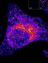
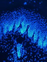
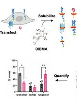
Members of the claudin family of tight junction proteins regulate paracellular permeability and modulate cell signaling. During junction remodeling, these proteins are selectively inserted into or retrieved from the tight junctions, but the control and coordination of these processes remain incompletely understood. Visualization of claudins allows the assessment of changes in their localization and abundance. We use the described protocol to stain claudin-2, but it can also be adapted to stain any tight junction protein. We found that using methanol for fixing allows the best preservation of claudin-2 both at the membrane and in cytoplasmic vesicles. Staining is done using a claudin-2 specific primary and a fluorescently labelled secondary antibody, along with DAPI to label nuclei. The samples are then imaged using confocal microscopy, and a z-stack is obtained allowing visualization of both junctional and intracellular claudin-2. Total claudin-2 signal can be quantified after 3D reconstruction of the images using the Imaris software.
Keywords: Claudin (Claudin)Background
Tight junctions (TJ) are multiprotein complexes localized at the apical-most portion of the intercellular junctional complexes that connect epithelial cells (Van Itallie and Anderson, 2014). These structures generate a permeability barrier and ion-specific paracellular pathways, maintain apico-basal polarity, provide input for various vital functions and modulate signaling pathways. The proteins located at the TJs can be divided into transmembrane and associated cytoplasmic proteins (reviewed in (González-Mariscal et al., 2003). The transmembrane proteins can be further subdivided into three major categories: claudins, tight junction-associated Marvel proteins (e.g., occludin) and single span proteins (e.g., Junctional Adhesion Molecules). TJ-associated cytosolic proteins include a large array of adaptors connected to signaling and cytoskeletal proteins. Collectively, these proteins are referred to as the cytoplasmic plaque. Claudins are small molecular weight (20-34 kDa) tetraspan membrane proteins that are essential components of the TJs (Tsukita et al., 2019). They incorporate into the TJ strands that typically contain a mosaic of various claudin isoforms that determine permselectivity (Van Itallie and Anderson, 2004). Interestingly, claudins have also recently emerged as key modulators of signaling, an effect that is likely due to the interactions with cytosolic adapters.
In this protocol we focus on claudin-2 (Cldn-2), a 230 amino acid member of the family, with a calculated molecular mass of 24.5 kDa. Cldn-2 was first described by Dr Shoichiro Tsukita and coworkers (Furuse et al., 1998a and 1998b) (for review see Venugopal et al., 2019). It is highly enriched in the kidney proximal tubules (Enck et al., 2001) and in intestinal and liver cells (e.g., Sakaguchi et al., 2002; Escaffit et al., 2005). It generates a cation selective paracellular channel, and therefore, its presence corresponds to elevated paracellular permeability (e.g., Amasheh et al., 2002). Multiple studies have revealed that in various cells Cldn-2 can be found not only at the membrane, i.e., in the TJs, but also in intracellular vesicles (Dukes et al., 2012; Lu et al., 2014; Amoozadeh et al., 2015), and in the nucleus (Ikari et al., 2014; Amoozadeh et al., 2018).
Many studies have used immunofluorescent staining in fixed cells followed by confocal microscopy to visualize TJ proteins and analyze their localization and abundance. These approaches allow investigation of the mechanisms that regulate TJ protein trafficking. The protocols differ in the fixation and blocking method and use primary antibodies targeted against specific proteins. Here we describe the method used in our lab to stain Cldn-2 in cultured kidney tubular cells based on (Amoozadeh et al., 2015 and 2018; Dan et al., 2019). We also detail our protocol for 3D reconstitution and analysis of changes in staining intensity following treatment, e.g. with cytokines. While we focus specifically on claudin-2, the same approach can be used for other TJ proteins in any epithelial or endothelial cells.
Materials and Reagents
- Micro cover glasses, round, No. 1, 18 mm (VWR® International, catalog number: CA-48380-046 ), sterilize coverglasses using autoclave
- Microscope slides (Fisherbrand, Fisher Scientific, catalog number: 12-550-15 )
- Clear tissue culture-treated 12-well microplates, (Corning Costar®, Millipore-Sigma, catalog number: CLS3513 )
- Parafilm
- Paper towel
- Cell line: LLC-PK1 kidney tubule epithelial cell line (European Collection of Authenticated Cell Cultures (Salisbury, UK), catalog number: 86121112 )
- For cell culture:
- Dulbecco's Modified Eagle Medium (D-MEM), low glucose, with L-glutamine
- 110 mg/L sodium pyruvate (Thermo Fisher, Gibco, catalog number: 11885084 )
- 10% Fetal Bovine Serum (FBS) (Thermo Fisher, Gibco, catalog number: 12483-020 )
- 1% Penicillin-Streptomycin (Penicillin-Streptomycin, 100x, sterile-filtered, cell culture tested) (Millipore-Sigma Aldrich, catalog number: P-4333 )
- Trypsin-EDTA (0.05% Trypsin with EDTA 4Na) (Thermo Fisher, Gibco, catalog number: 25300062 )
- Phosphate buffered saline (PBS) (Thermo Fisher, Gibco, catalog number: 10010023 )
- Dulbecco's Modified Eagle Medium (D-MEM), low glucose, with L-glutamine
- Methanol (BioShop Canada, catalog number: Met302 ), cool to -20 °C before using
- 10x PBS Buffer (pH 7.4 Sterile, w/o Ca, Mg) (BioShop Canada, catalog number: PBS415 ), open in hood to maintain sterility for storage
- Bovine serum albumin (BSA) (fraction V, heat shock isolation, > 98%) (BioShop Canada, catalog number: ALB001 )
- Polyclonal rabbit anti-Claudin-2 antibody (Thermo Fisher Scientific, Invitrogen, catalog number: 28530 ), aliquot to avoid repeated thawing, store at -20 °C
- Fluorophore-labeled secondary antibody:
Donkey anti-Rabbit IgG labelled with Alexa Fluor 488 (green) (Thermo Fischer Scientific, catalog number: A-21206 )
or
Donkey anti-Rabbit IgG labelled with Alexa Fluor 555 (red) (Thermo Fischer Scientific, catalog number: A- A31572 ) - 2,4-diamidino-2-phenylindole (DAPI) (Thermo Fischer Scientific, catalog number: D1306 )
Make a 5 mg/ml DAPI stock solution by dissolving the contents of one vial (10 mg) in 2 ml of deionized water. Aliquot the stock, protect from light. DAPI stock solution can be stored at 4 °C for up to 6 months, or at -20 °C for longer periods. - Fluorescent mounting medium (Dako, Agilent catalog number: S3023 ), store at 4 °C
- Cell culture medium (see Recipes)
- 1x PBS (see Recipes)
- Blocking buffer (see Recipes)
- Antibody buffer (see Recipes)
Equipment
- Tweezers
- Nikon Eclipse TS100 microscope
- WaveFX spinning-disk confocal microscope (Quorum Technologies, Guelph, Canada) with an ORCA-flash4.0 digital camera with Gen II sCMOS image sensor
Software
- Metamorph Image Analysis Software, Molecular Devices
- Imaris software 8.0.2 (Bitplane)
Procedure
文章信息
版权信息
© 2020 The Authors; exclusive licensee Bio-protocol LLC.
如何引用
Readers should cite both the Bio-protocol article and the original research article where this protocol was used:
- Anwer, S. and Szászi, K. (2020). Immunofluorescent Staining of Claudin-2 in Cultured Kidney Tubular Cells. Bio-protocol 10(14): e3678. DOI: 10.21769/BioProtoc.3678.
- Dan, Q., Shi, Y., Rabani, R., Venugopal, S., Xiao, J., Anwer, S., Ding, M., Speight, P., Pan, W., Alexander, R. T., Kapus, A. and Szászi, K. (2019). Claudin-2 suppresses GEF-H1, RHOA, and MRTF, thereby impacting proliferation and profibrotic phenotype of tubular cells. J Biol Chem 294(42): 15446-15465.
分类
生物化学 > 蛋白质 > 荧光
癌症生物学 > 炎症 > 细胞生物学试验
细胞生物学 > 细胞染色 > 蛋白质
您对这篇实验方法有问题吗?
在此处发布您的问题,我们将邀请本文作者来回答。同时,我们会将您的问题发布到Bio-protocol Exchange,以便寻求社区成员的帮助。