

Precise Targeting of Single Microelectrodes to Orientation Pinwheel Centers
单微电极精确定位风车中心区

(*contributed equally to this work) 发布: 2020年06月05日第10卷第11期 DOI: 10.21769/BioProtoc.3643 浏览次数: 3947
评审: Miao HeEhsan KheradpezhouhAnonymous reviewer(s)
参见作者原研究论文
The authors used this protocol in:

Jun 2019


Abstract
In the mammalian visual system, early stages of visual form perception begin with orientation selective neurons in primary visual cortex (V1). In many species (including humans, monkeys, tree shrews, cats, and ferrets), these neurons are organized in pinwheel-like orientation columns. To study the functional organization within orientation pinwheels, it is important to target pinwheel subdomains precisely. We therefore developed a technique to provide a quantitative determination of the location of pinwheel centers (PCs). Previous studies relied solely on blood vessel images of the cortical surface to guide electrode penetrations to PCs in orientation maps. However, considerable spatial error remained using this method. In the present study, we improved the accuracy of targeting PCs by ensuring perpendicularity of electrodes and by utilizing the orientation tuning of local field potentials (LFP) recorded at or near the optically determined positions.
Keywords: Orientation domain (方向功能区)Background
Within the visual cortex, orientation pinwheels (PCs) are singularity centers around which neuronal orientation preference systematically shifts. PCs have been the focus of many studies on how the brain encodes different aspects of object shape, such as linear oriented segments, curved segments, corners, and T junctions (Hubel and Wiesel, 1974; Das and Gilbert, 1999; Hashemi-Nezhad and Lyon, 2012). Single-unit recording has been the most common method for studying organization within orientation pinwheels (Hubel and Wiesel, 1974). However, due to limitations of blood vessel guided localization of microelectrodes and uncertainties of electrode perpendicularity (Nauhaus and Ringach, 2007), it is quite challenging to accurately target microelectrodes to pinwheel centers (PCs). Hashemi-Nezhad and Lyon (2012) used electrodes coated with Dil to nicely demonstrate that electrodes were truly perpendicular; however, the vasculature-based method still contained localization error. Nauhaus and Ringach (2007), carefully matched the electrode locations in a Utah array with locations in an optical imaged orientation map and achieved accurate PC localization, but such an approach offers little control over where the electrodes end up within orientation domains or pinwheel centers. Using 2-photon methods, Ohki et al. (2006) reported that the true PC is only 130 µm in diameter, underscoring the need for highly accurate electrode targeting (see also Nauhaus et al., 2008).
In this study, we developed a method for targeting PCs with high accuracy. This was achieved by: (1) Assuring perpendicular penetrations by imaging a large field of view with a narrow depth of field (~50 μm, front-to-front lens with f1.2 (Ratzlaff and Grinvald, 1991), and then ensuring the electrode paralleled exactly the axis of the optical imaging. Using this procedure, we have estimated that the deviation of electrodes from perpendicular is less than 2 deg (see Figure 3D). (2) Carefully targeting electrodes to PCs based on blood vessel guidance. (3) Further determining the precise PC center by assessing whether the local field potentials (LFP) exhibit ‘non-sine-like’ responses. We have previously shown (Li et al., 2019) using this methodology that penetrations with sine-like LFP response and small scatter in orientation preference are not true PC locations, while those with non-sine-like response and large scatter in orientation preference (> 60 deg, Maldonado et al., 1997) are the true PC locations. This additional non-sine-like criterion is essential for highly accurate determination of PC locations and has provided new understanding of orientation subdomain organization (Li et al., 2019). These detailed procedures are described below.
Materials and Reagents
- Cover glass
- Tungsten-in-glass microelectrodes (impedance: ~5 MΩ; tip diameter: ~1 μm):
These electrodes were homemade. Detailed description of procedures for electrode construction can be found in previous paper (Li et al., 1995). Briefly, tungsten-in-glass electrodes were prepared from tungsten wire (diameter 200 µm, Shanghai Tungsten and molybdenum products company, type: S-EST6135) and capillary glass (inner diameter: 0.3 mm, outer diameter: 1 mm, borosilicate glass, Shanghai Glass Works, type: SGW-95). The desired impedance of ~5 MΩ was measured (Ωmega-Tip-Z, World Precision Instruments,USA ) and corresponded to tungsten tip length of ~10 µm and the tip diameter of ~1 µm (Figure 1).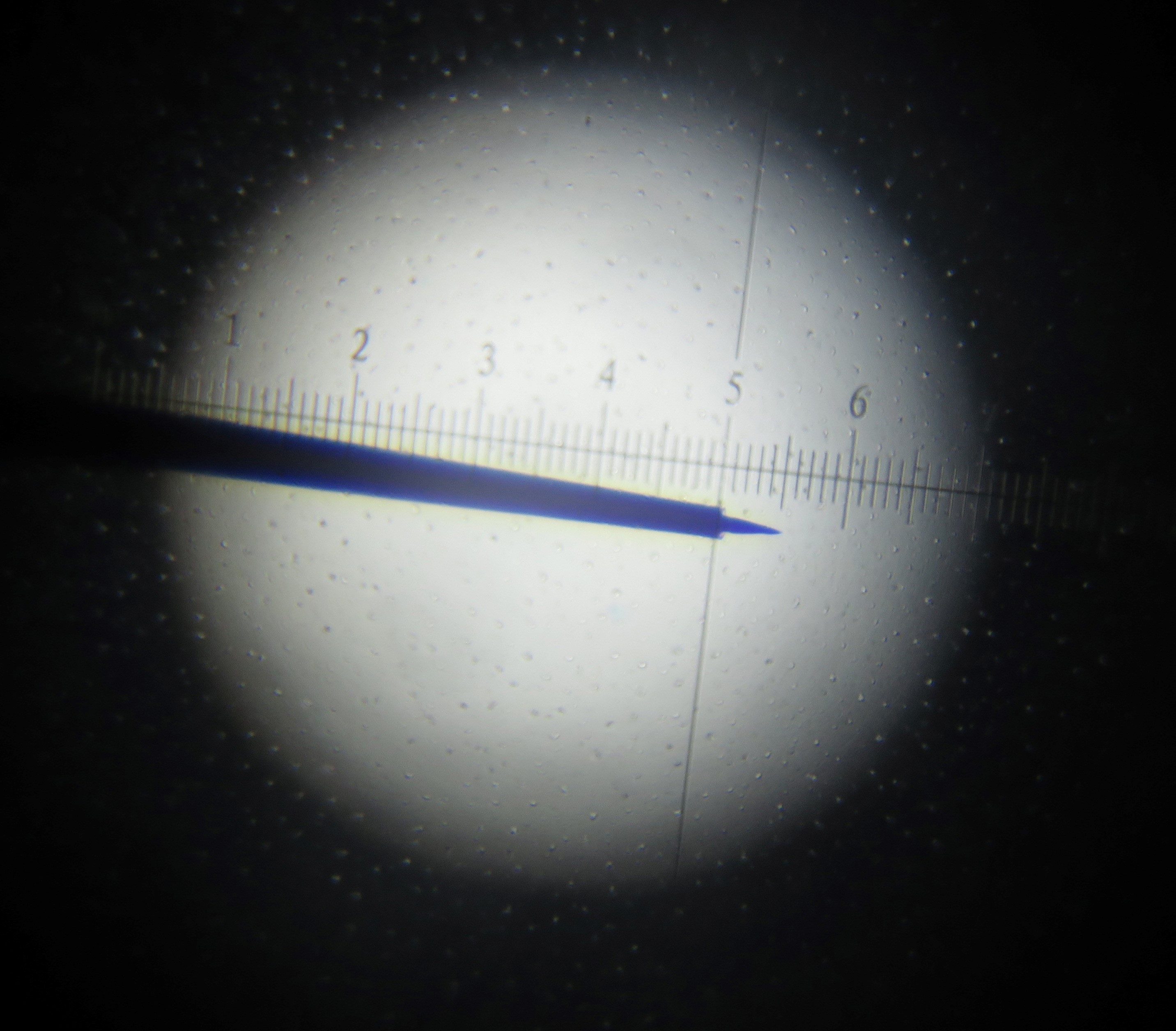
Figure 1. Photograph of tungsten-in-glass microelectrode. 1 grid mark is 2.5 µm Image shows electrode tip length of of ~10 µm, and tip diameter of ~1 µm. - Animals
These procedures were developed using 16 anesthetized adult cats of both sexes. The following procedures can be applied to the visual cortex of other species as well. - Ketamine hydrochloride
- 1% atropine sulfate eyedrops
- Agar
- Barbiturate
Equipment
- Neural data acquisition system (Blackrock Microsystems Inc., Cerebus, Cerebus 32)
- Video camera (Andor Technology, Andor, model: iXon DU897 )
- Lenses (Nikon, NIKKOR, D = 50 mm, f = 1.2)
- Visual stimulus Generator (Cambridge Research Systems Ltd. ViSaGe MKII)
- Stereotaxic apparatus (Narishige, SN-3N)
Software
- Matlab (Mathworks, ww2.mathworks.cn), and the matlab code as the supplemental file
Procedure
文章信息
版权信息
© 2020 The Authors; exclusive licensee Bio-protocol LLC.
如何引用
Readers should cite both the Bio-protocol article and the original research article where this protocol was used:
- Song, X. M., Li, M., Xu, T., Hu, D. and Roe, A. W. (2020). Precise Targeting of Single Microelectrodes to Orientation Pinwheel Centers. Bio-protocol 10(11): e3643. DOI: 10.21769/BioProtoc.3643.
- Li, M., Song, X. M., Xu, T., Hu, D., Roe, A. W. and Li, C. Y. (2019). Subdomains within orientation columns of primary visual cortex. Science Advances, eaaw0807.
分类
神经科学 > 感觉和运动系统 > 视觉系统
神经科学 > 感觉和运动系统 > 动物模型
您对这篇实验方法有问题吗?
在此处发布您的问题,我们将邀请本文作者来回答。同时,我们会将您的问题发布到Bio-protocol Exchange,以便寻求社区成员的帮助。