

Isolating Multiple Extracellular Vesicles Subsets, Including Exosomes and Membrane Vesicles, from Bovine Milk Using Sodium Citrate and Differential Ultracentrifugation
用柠檬酸钠和差速超速离心分离牛乳中多个含外泌体和膜囊泡的胞外小泡亚群

发布: 2020年06月05日第10卷第11期 DOI: 10.21769/BioProtoc.3636 浏览次数: 6679
评审: Karem A CourtAlexandros C KokotosAnonymous reviewer(s)
参见作者原研究论文
The authors used this protocol in:

Oct 2019
Advertisement


Abstract
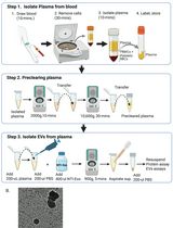
Milk is a complex fluid that contains various types of proteins and extracellular vesicles (EVs). Some proteins can mingle with EVs, and interfere with their isolation. Among these proteins, caseins form micelles of a size comparable to milk EVs, and can thus be co-isolated with EVs. Preliminary steps that affect milk are crucial for EV isolation and impact the purity and abundance of isolated EVs. In the course of our previous works on cow’s milk EVs, we found that sodium citrate (1% final), which is a biocompatible reagent capable of breaking down casein micelles into 40-nm monomers, allowed the isolation of high quantities of EVs with low coprecipitation of caseins or other contaminating proteins. Using this protocol, we successfully separated different EV subsets, characterized in depth their morphology, protein content and small RNA enrichment patterns. We were also able to describe their biological function in a mouse model of intestinal inflammation. We, hereby, detail the differential ultracentrifugation procedure that leads to high quantify, medium specificity, isolation of different milk EV subsets from the same sample. More specifically, we highlight the use of sodium citrate as a standardized approach to isolate and study milk EVs and its potential for isolation techniques other than differential ultracentrifugation.
Keywords: Extracellular Vesicles (细胞外基质)Background
In our previous publications (Benmoussa et al., 2016, 2017, 2019b and 2019c; Benmoussa and Provost, 2019), we highlighted that bovine milk is a complex fluid containing a myriad of extracellular vesicles (EVs) subsets. Among these, exosomes are ~100-nm vesicles released when multivesicular bodies (MVB) fuse with the cell membrane. When subjected to ultracentrifugation, these sediment at centrifugation speeds equal or higher than 100,000 x g (P100K, where P stands for pellet) (Pieters et al., 2015). Other non-exosome EV subsets are found in milk and are comparable to exosomes, in shape and size, but sediments at a lower speed (e.g., 12,000 x g, P35K; 35,000 x g, P35K; 70,000 x g, P100K). These are thought to be originating from budding of the cell membrane and contain specific proteins and microRNAs different from exosomes content (Benmoussa et al., 2016, 2017, 2019b and 2019c; Benmoussa and Provost, 2019).
For a long time, separating these different milk EVs subsets remained challenging because of their morphological and chemical similarities. Moreover, protocols used in milk EV studies were often centered on the isolation of milk exosomes specifically, leading to the discarding of the biologically active non-exosome milk EVs (Benmoussa et al., 2019a, 2019b and 2019c).
When using differential ultracentrifugation to isolate milk EVs, the first steps are often impaired by the co-precipitation of milk proteins with EVs, which form a dense jelly at the bottom of the tubes (Zonneveld et al., 2014). Such jelly is formed when caseins are subjected to high mechanical pressures (Famelart et al., 1998; Zonneveld et al., 2014). Because this jelly is so dense, and practically impossible to resuspend, it is not clear whether some EV subsets are trapped within its matrix and some protocols recommend discarding this casein-rich jelly.
Certain reports suggested the use of density gradients or cushions to keep the EVs from mixing with the casein jelly (Zonneveld et al., 2014). Others suggested avoiding the formation of such jelly by discarding caseins before subjecting milk “serum” or whey to differential ultracentrifugation. To this end, milk is often mixed with acids (Somiya et al., 2018) or with cold EDTA to precipitate the caseins prior to milk EVs sedimentation (Wolf et al., 2015). However, such preprocessing of biological fluids have an immense impact on the isolation, quality and yield of extracellular vesicles (EVs) (Zonneveld et al., 2014). It also requires to discard low speed-pelleting EVs (P12K or P35K). In addition, there is a lack of information about the effect of acidification, or casein precipitation, on milk EVs, and the possibility that some EVs might be lost along the process.
This is especially important because there are multiple EV subsets in milk (Benmoussa et al., 2017 and 2019b) and because milk whey has a different microRNA and protein content than milk (Benmoussa et al., 2019b; Benmoussa and Provost, 2019), which, as microRNA are found within EVs, suggests the loss of certain EVs during casein precipitation. This is also supported by the discovery of microRNAs in milk-derived casein-rich products, like cheese (Benmoussa and Provost, 2019). Therefore, the biological activity of the milk EVs, and the bioactive molecules they transport, depend highly on the reagents/preprocessing steps and isolation protocols this fluid is exposed or subjected to (Zonneveld et al., 2014).
When we encountered this issue, we experimented different venues to avoid precipitating milk caseins while ensuring the separation of different structurally conserved, and functional, milk EV subsets. In milk, casein proteins are arranged in the form of micelles that are 70 to 200 nm in diameter, which is close to the size of certain EV subsets (Blans et al., 2017). Knowing that calcium is important for maintaining these micelles (de Kort et al., 2009, 2011 and 2012; Kort et al., 2012), we supposed that calcium chelation would prevent the formation of casein superstructures that hamper EV isolation by ultracentrifugation.
Several calcium-chelating agents have been previously investigated for their ability to disrupt casein micelles, including disodium uridine monophosphate (Na2UMP), disodium phosphate (Na2HPO4), trisodium citrate (referred to simply as "sodium citrate"), sodium phytate, sodium hexametaphosphate (SHMP) or ethylenediaminetetraacetic acid (EDTA) (Ward et al., 1997; de Kort et al., 2009, 2011 and 2012; Kort et al., 2012). These chelating agents interact with colloidal calcium phosphate (CCP) leading to the breaking of casein micelles into small monomers. This process augments the stability of milk during heating and change its physical properties, often reducing its viscosity (de Kort et al., 2009, 2011 and 2012; Kort et al., 2012).
Among these, sodium citrate is a biocompatible compound known for 100+ years for its ability to prevent casein curdling in the stomach of infants (Poynton, 1904). It is widely used to preserve biological fluids, like blood (Janse van Rensburg and van der Merwe, 2017), as a food additive and as the major form of rehydration salt recommended by the World Health Organization (Pizarro et al., 1986; Banipal et al., 2016). Notably, it was previously used to disrupt casein micelles to facilitate the isolation of milk fat globules (MFGs) by diafiltration and prevented pore clogging (Phan et al., 2014). Interestingly, sodium citrate specifically impacts milk protein gel formation upon high mechanical pressure, although the underlying mechanisms remain unclear, except for its dependence on the solution’s pH (Famelart et al., 1998).
In the course of our experimentations with sodium citrate, we found that pre-treating milk with sodium citrate (1% final) was a cost-effective way to avoid casein jellification and isolation of high quantities of different milk EVs using differential ultracentrifugation. We hereby detail the methods underlying this method that led to the discovery (Benmoussa et al., 2016 and 2017) and characterization (Benmoussa et al., 2017, 2019b and 2019c) of different EVs subsets in milk, keeping them functional and able to transfer their content to human cells (Benmoussa et al., 2019c). EVs, including exosomes, isolated by this methodology were able to modulate intestinal inflammation during experimental colitis (Benmoussa et al., 2019a).
Materials and Reagents
- Sterilized glass bottle (Pyrex, Corning Life-Science, catalog number: 1395-1L) or sterile plastic bottles for mixing milk and sodium citrate (Corning, catalog number: 431533)
- Falcon tubes 50 ml (any product, as long as sterile and pyrogen-free, e.g., Corning, catalog number: 352070, Fisher Scientific, catalog number: 14-432-22)
- 1.7 ml snap cap tubes (any product as long as sterile and pyrogen-free, e.g., Corning, Costar via Sigma-Aldrich, catalog number: CLS3620)
- 0.22 µm membrane microfilters (Corning, Sigma-Aldrich, catalog number: CLS431224)
- Serological pipets different volumes (any product as long as sterile and pyrogen-free, e.g., Sigma-Aldrich, catalog number: SIAL1485)
- Filtered 1 ml pipet tips (any product as long as sterile, pyrogen free and filtered, e.g., Thermo Scientific, catalog number: 94052410)
- Nitrile disposable gloves (any type)
- Sterile wipes/gauze (any as long as sterile single-packed, e.g., VWR, catalog number: CA95041-740)
- Optional: EDTA. Can be bought as ready-to-use solution (Sigma-Millipore, catalog number: 324506) or homemade from powdered EDTA
- Milk
Use commercially available milk bought on the day of the experiment or raw cow milk collected and stored in “sterile conditions” (as sterile as possible using sterilized bottles and keeping the bottles closed until getting under a biological hood). In our work we used mostly pasteurized commercial ultrafiltered skim milk (Lactantia PureFilter, bought in a local grocery store) as it avoids creaming steps that could lead to variability. We recommend working with a pool of three milks with different preemption dates. Milk must be stored at 4 °C.
Note: If working with raw milk, use pool from different cows to minimize interindividual differences. Noncommercial raw or pasteurized fresh cow milk can be frozen at -80 °C to avoid bacterial contamination/proliferation but note such storage might impact EVs content. If freezing milk, make sure it is previously skimmed and decellularized to minimize EVs formations due to milk fat globule or cell destruction upon freeze/thawing cycles. Freeze milk in small aliquots (50 ml) so, when thawing, it reaches melting point quicker and limit EVs destruction. - Bradford's reagent (Sigma-Aldrich, catalog number: B6916)
- BCA Protein Assay Kit (Millipore, catalog number: 71285-M)
- Milli-Q water or any filtered ultra-pure water (e.g., Millipore, water SystemClear Sorting & Filtering, catalog number: ZRXQ003WW)
- NaCl (preferably low in endotoxins, e.g., Millipore-Sigma, SAFC, catalog number: 1.16224)
- KCl (e.g., Sigma-Aldrich, catalog number: P3911)
- Na2HPO4 (e.g., Sigma-Aldrich, catalog number: NIST2186II)
- KH2PO4 (e.g., Sigma-Aldrich, catalog number: NIST200B)
- HCl (e.g., Sigma-Aldrich, catalog number: 320331)
- EDTA (e.g., Sigma-Aldrich, catalog number: EDS-500G)
- NaOH solution in Milli-Q water (e.g., Sigma-Aldrich, catalog number: S8045)
- Sodium citrate dihydrate (Sigma-Aldrich, catalog number: W302600)
- Phosphate buffered saline
Either bought as commercially available as filtered sterile phosphate buffered saline (PBS) pH 7.4 (Sigma-Aldrich, catalog number: P5493) or homemade. - 2% sodium citrate solution (1 L) (see Recipes)
- Phosphate buffer saline (PBS) 10x (1 L) (see Recipes)
- EDTA 0.5 M recipe (100 ml) (see Recipes)
Equipment
- Tubes for ultracentrifugation compatible with rotor bellow (36 ml, Beckman, catalog number: 355631 or 344058 or other, see rotor compatibility)
- Ultracentrifuge Sorvall WX ultracentrifuge (or one of Thermo ScientificTM SorvallTM WX ultraCentrifuges, catalog numbers: 75000100, 7500090, 7500080)
- Rotor SureSpin 630 swinging bucket Rotor (Thermo Scientific, catalog number: 79368)
Note: You can use another centrifuge and another rotor but make sure to get comparable material. Note that it is not recommended to use fixed-angle rotors as they will induce higher EV degradation. - Pipetting device (any type, e.g., Drummond Scientific Portable Pipet-Aid XP via Mandel, catalog number: DRU-4-000-101)
- Orbital shaker (any type, e.g., Bel-ArtTM SP SciencewareTM SpindriveTM Orbital Shaker Platform, via Fisher-scientific, catalog number: 1451176)
- Rotating mixer/tube revolver (any type, e.g., Thermo-scientific, catalog number: 88881001)
- High precision balance (any type, e.g., Sartorius, catalog number: PRACTUM224-1S)
- Tube rack holder different sizes (any product)
- Laminar flow hood (if working on sterile conditions) (any product)
- Micropipettes, different volumes (any type as long as they are precise and pipetting is smooth enough to avoid too fast ejection of suspension liquid, e.g., PIPETMAN L P1000L, 100-1,000 µl, Metal Ejector, Gilson, catalog number: FA10006M)
- -80 °C freezer
- Magnetic stirrer
Procedure
文章信息
版权信息
© 2020 The Authors; exclusive licensee Bio-protocol LLC.
如何引用
Benmoussa, A., Michel, S., Gilbert, C. and Provost, P. (2020). Isolating Multiple Extracellular Vesicles Subsets, Including Exosomes and Membrane Vesicles, from Bovine Milk Using Sodium Citrate and Differential Ultracentrifugation. Bio-protocol 10(11): e3636. DOI: 10.21769/BioProtoc.3636.
分类
生物化学 > 蛋白质 > 分离和纯化
细胞生物学 > 细胞器分离 > 胞外囊泡
细胞生物学 > 细胞器分离 > 外来体
您对这篇实验方法有问题吗?
在此处发布您的问题,我们将邀请本文作者来回答。同时,我们会将您的问题发布到Bio-protocol Exchange,以便寻求社区成员的帮助。